Search results (309 results)
-
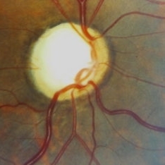
 OCT RNFL
-
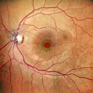
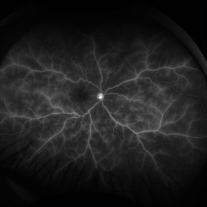
 OCT Fundus
Jul 26 2025 by oren reuven
The same patient with glaucoma and preserved fundus.
Condition/keywords: fundus of glaucoma paitiant
-
 Synechiae
Jul 26 2025 by oren reuven
Uveitis causing Synechiae leading to Glaucoma in a 74 year-old male.
Photographer: oren reuven
Condition/keywords: synechiae
-
 Synechiae
Jul 26 2025 by oren reuven
Uveitis causing Synechiae leading to Glaucoma in a 74 year-old male.
Photographer: oren reuven
Condition/keywords: synechiae
-
 Synechiae
Jul 26 2025 by oren reuven
Uveitis causing Synechiae leading to Glaucoma in a 74 year-old male.
Photographer: oren reuven
Condition/keywords: synechiae
-
 Synechiae
Jul 26 2025 by oren reuven
Uveitis causing Synechiae leading to Glaucoma in a 74 year-old male.
Photographer: oren reuven
Condition/keywords: synechiae
-
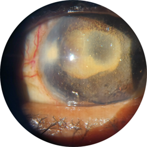
 Synechiae
Jul 26 2025 by oren reuven
74 year-old male, 4 years of uveitis casing Synechiae that closed the chamber angle and caused glaucoma. Suffering minor field loss
Photographer: oren reuven
Imaging device: EIDON
Condition/keywords: glaucoma and uveitis
-
 Synechiae
Jul 26 2025 by oren reuven
74 year-old male, 4 years of uveitis casing Synechiae that closed the chamber angle and caused glaucoma. Suffering minor field loss.
Photographer: oren reuven
Imaging device: EIDON
Condition/keywords: synechiae, uveitis
-
 Rubeosis Iridis
Jul 21 2025 by Luai Abu-Ismail, MD
73y old female patient, known case of uncontrolled DM for more than 15y and chronic kidney disease. She had CRVO and complicated with 100 day neovascularization glaucoma.
Photographer: Luai Abu-Ismail
Imaging device: S23 Ultra
Condition/keywords: central retinal vein occlusion (CRVO), neovascular glaucoma, Rubeosis
-
 Double Trouble
Jun 28 2025 by Tejaswita Verma
Fundus image of a 60 year old diabetic female with double macular holes with 6/60 vision status post LE PPV+gas 4 months ago. Other eye also had an unoperated large macular hole. Known case of glaucoma
Photographer: Dr. Tejaswita Verma
Imaging device: MIRANTE
Condition/keywords: macular hole
-
 Multimodal Imaging in CHRPE
Mar 6 2025 by Gerardo - Montante Montelongo, MD
Fundus photograph of an 83-year-old male with a history of Diabetes, smoking, cataract surgery on the right eye in 2022, and open-angle glaucoma. Asymptomatic. Indirect ophthalmoscopy revealed 80% excavation, peripapillary atrophy, and a hyperpigmented perifoveal lesion with 35% atrophy, 10% drusen, and 5.1 mm diameter, corresponding to a CHRPE. At multimodal imaging, FFA shows hypoautofluorescence of the lesion, OCT shows preservation of internal retinal layers, atrophy of external retinal layer, with an RPE disruption, and posterior shadowing. USG shows a flat hyperechoic lesion 5.1 mm in diameter and 1.32 mm in thickness, solid and with high internal reflectance.
Photographer: Gerardo Montante-Montelongo, MD, Mexican Institute of Ophthalmology
Imaging device: Clarus 700
Condition/keywords: congenital hypertrophy of the retinal pigment epithelium (CHRPE), multimodal imaging
-
 Firework Injury
Feb 13 2025 by Virginia Gebhart
44 year old male presented New Year's Day for trauma after fireworks injury. Choroidal rupture temporal macula, inferior vitreous hemorrhage, and extensive RPE changes in the macula. Significant improvement since initial presentation. Limited central vision, guarded prognosis due to extensive blunt trauma.
Photographer: Virginia Gebhart, Retina Consultants of Carolina
Imaging device: Optos California
Condition/keywords: blunt trauma, choroidal rupture, commotio retinae, firework injury, secondary glaucoma, subretinal hemorrhage, VH, vitreous hemorrhage
-
 Retinal Arteriolar Variation
Oct 31 2024 by AVIK DEY SARKAR, MS, FVRS, FAICO(VR)
A 43-year-old hypertensive patient, diagnosed with Non-Ischemic Central retinal vein Occlusion in OS, presented with a striking anatomical variation in retinal vasculature. The inferior first-order retinal arteriole after initiating from the optic disc bifurcates, before reaching the fovea, and the superior branch after crossing the midline forms the superior arcade afterwards and produces dichotomous branching as usual. This defies basic anatomical considerations for retinal vasculature as they never cross the midline, also known as the watershed line for retinal vessels.1,2 References: 1. May CA, Rutkowski P. The Horizontal Raphe of the Human Retina and its Watershed Zones. Vision. 2019; 3(4):60. 2. May CA, Rutkowski P. Hypothesis: watershed zones in the human eye are a key for understanding glaucomatous retinal damage. Med Hypotheses. 2017;109:1-5.
Photographer: Dr. Avik Dey Sarkar, MBBS, MS, FVRS, FAICO, Consultant, Department of Vitreoretinal Services, Aravind Eye Hospital, Madurai, India
Imaging device: Wide angled Fundus imaging with Clarus 300
Condition/keywords: background diabetic retinopathy (BDR), Diabetic Retinopathy, retina, vascular anomaly
-
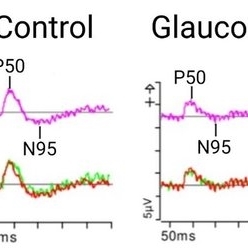 Representative Pattern Electroretinography Responses
May 13 2024 by Gabrielle Hallai
The pattern ERG response on the left is from a control individual with no known retinal pathology. There is a clearly discernable P50 and N95 peak. On the right, there is a representative image from a patient with end-stage glaucoma. In advanced glaucoma, there is often a lack of a clearly discernable N95 peak along with a diminished N95:P50 amplitude ratio, due to retinal ganglion cell dysfunction or degeneration. The top traces are averages from two independent trials shown on the bottom. Pattern ERG testing was completed using the Diagnosys pattern ERG protocol on a CRT monitor.
Photographer: Gabrielle Hallai, PhD, Cleveland Clinic Cole Eye Institute
Condition/keywords: electroretinography, glaucoma, pattern ERG (PERG)
-
 Crystallized silicone oil particles in the anterior chamber
Oct 26 2023 by Anmol Naik, MS, DNB, FMRF, FICO, MNAMS
Anterior segment image of a 67-year-old Indian woman who had proliferative diabetic retinopathy with traction retinal detachment with neovascular glaucoma. Patient underwent vitrectomy with membrane peeling with endolaser followed by silicone oil injection 1 year back. Patient was lost to follow up and presented a year later with this picture. She had crystallized silicone oil particles in the anterior chamber rendering a polychromatic lustre like appearance; a unique and rare finding.
Photographer: Anmol Naik
Condition/keywords: Polychromatic lustre in Anterior Chamber
-
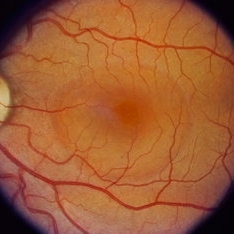
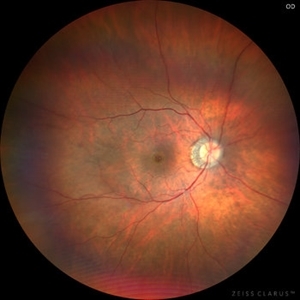
 Glaucomatous Optic disc cupping
Sep 21 2023 by Ben Serar
Fundus photograph showing increased cup-disc ratio with nasalisation of vessels in a case of Glaucoma.
Condition/keywords: Glaucomatous Optic disc cupping
-
 Central Serous Chorioretinopathy (CSR)
Sep 21 2023 by Ben Serar
Fundus photograph showing increased cup-disc ratio with nasalisation of vessels , with thinning of Neuroretinal rim and bayonetting of vessels in a case of Glaucomatous Optic Atrophy (GOA) Fundus photograph of LE showing serous macular detachment in a case of Central Serous Chorioretinopathy (CSR).
Condition/keywords: Central Serous Chorioretinopathy (CSR)
-
 Glaucomatous Optic Atrophy (GOA)
Sep 21 2023 by Ben Serar
Fundus photograph showing increased cup-disc ratio with nasalisation of vessels , with thinning of Neuroretinal rim and bayonetting of vessels in a case of Glaucomatous Optic Atrophy (GOA).
Condition/keywords: Glaucomatous Optic Atrophy (GOA)
-
 Glaucomatous Optic disc cupping
Sep 14 2023 by Ben Serar
Fundus photograph showing increased cup-disc ratio with nasalisation of vessels in a case of glaucoma.
Condition/keywords: Glaucomatous Optic disc cupping
-
 Glaucomatous Optic Atrophy (GOA)
Sep 12 2023 by Ben Serar
Fundus photograph of the LE showing enlarged optic cup with disc pallor in a case of Glaucomatous Optic Atrophy (GOA)
Condition/keywords: Glaucomatous Optic Atrophy, GOA
-
 Retinal Vasculitis
Aug 22 2023 by Karen Santamaría
Fluorescein angiography of patient of a 23 year-old man diagnosed with retinal vasculitis and juvenile glaucoma.
Photographer: Karen Santamaría, Hospital Militar de Especialidades Oftalmológicas - Servicio de Glaucoma, Ciudad de México
Imaging device: Optos California
Condition/keywords: juvenile glaucoma, retina, vasculitis
-
 Advanced Glaucoma
Aug 21 2023 by Harsh Vardhan Singh, MS
A 64-year-old female with glaucomatous changes in both eye with advanced glaucoma in left eye
Photographer: Harsh Vardhan Singh
Condition/keywords: advanced stage glaucoma, Asymmetric cupping, glaucoma
-
 Limited Choroidal Hemorrhage S/P Glaucoma Valve Implant OS; Retinoschisis
Aug 21 2023 by Angela Rico
A 52 year old Female presents to office S/P Glaucoma Valve Implant with IOP: 5mmHg OS
Photographer: Angela Rico M.D.
Condition/keywords: choroidal hemorrhage, glaucoma, hypotony, retinoschisis
-
 Central Retinal Vein Occlusion
Aug 17 2023 by Karen Santamaría
Fundus photograph of a female patient with a disc neovascularization secondary to central retinal vein occlusion.
Photographer: Karen Santamaría, Hospital Militar de Especialidades Oftalmológicas - Servicio de glaucoma, Ciudad de México
Imaging device: OCT RS-330 Retina Scan Duo 2 - NIDEK
Condition/keywords: Central vein oclussion, glaucoma
-
 Glaucoma with full thickness macular hole
Jul 31 2023 by Harsh Vardhan Singh, MS
58-year-old male with open angle glaucoma & full thickness macular hole
Photographer: Dr Harsh Vardhan Singh, AIIMS, Guwahati
Imaging device: Zeiss Clarus 700
Condition/keywords: full thickness macular hole, glaucoma
 Loading…