Search results (1109 results)
-
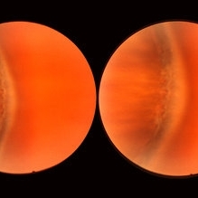
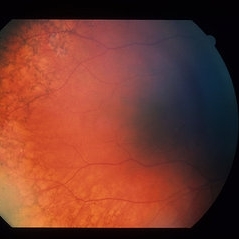
 Lacquer Cracks
Oct 13 2012 by Geoffrey G. Emerson, MD, PhD, FASRS
Lacquer cracks
Condition/keywords: lacquer cracks, myopic macular degeneration
-
 Normal Temporal Ora Serrata
Nov 9 2012 by Norman Byer
This is the normal temporal ora serrata in a 26-year-old man. Note the typical ragged moth-eaten appearance caused by peripheral cystoid degeneration. This appearance may be present in infants but is always present beyond the age of eight years.
Condition/keywords: ora serrata, peripheral cystoid degeneration
-
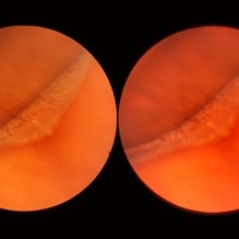
 Meridional Fold
Nov 9 2012 by Norman Byer
This is the same lesion as in the previous photograph. With the scleral indentation placed more posterior, we now can see that the fold ends over a small collection of subretinal fluid and that there is a very tiny retinal hole just below the posterior end of the retinal fold.
Condition/keywords: peripheral cystoid degeneration, retinal fold, retinal hole, scleral indentation, subretinal fluid
-
 Operculated Retinal Hole
Oct 12 2012 by Jeffrey G. Gross, MD, FASRS
Operculated retinal hole with RD.
Condition/keywords: operculated retinal hole, retinal degeneration
-
 Lattice Degeneration
Nov 9 2012 by Norman Byer
This is a more typical classical example of lattice degeneration in a 42-year-old woman in a photograph taken without scleral indentation. It shows much more marked vascular changes than the previous case. Note the tapering of the blood columns as the vessels approach the lesion and also the white sheathing of the vessel walls. Note also the continuity of the blood vessels on opposite sides of the lesion with the characteristic white lattice lines. More than 45 years ago Vogt pointed this out as a proof that these white lines were actually caused by changed blood vessels. Note also that this lesion shows a combination of several individual features of lattice degeneration. In addition to the white lines, there is a reddish crater-like area beneath the main horizontal white line. There is a prominent horizontal zone below this white line showing a snailtrack appearance. Also, there are two tiny atrophic retinal holes outside the photograph on the right end of this lesion. This eye contained five such retinal holes and they have all remained unchanged for more than 10 years of observation without treatment.
Condition/keywords: atrophic retinal hole, lattice degeneration, moderate snail track, tapering blood columns, white lattice lines, white sheath vessel
-
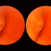
 Meridional Fold
Nov 9 2012 by Norman Byer
The next two photographs are of the same lesion in a 28-year-old woman. This view shows a sloping retinal mound with a radial retinal fold in the center. This is not a typical meridional fold for it stops short of the ora serrata and there is no dentate process. The upper temporal ora serrata and pars plana are well shown and peripheral cystoid degeneration is present posterior to the ora.
Condition/keywords: ora serrata, pars plana, peripheral cystoid degeneration, radial retinal fold, sloping retinal mound
-
---thumb.jpg/image-square;max$300,300.ImageHandler) Lattice Degeneration
Lattice Degeneration
-
 Lattice Degeneration
Nov 9 2012 by Norman Byer
This 16-year-old girl has lattice degeneration and also this large oval retinal hole with a surrounding narrow zone of subretinal fluid. This lesion illustrates how large the atrophic holes of lattice degeneration may be. Occasionally the hole can be as large as the initial lattice lesion and can therefore obliterate all other evidence of its true identity. This was almost true in this case, but there does remain a small whitish remnant of the original lattice lesion at the lower end of the oval hole.
Condition/keywords: lattice degeneration, retinal hole, subretinal fluid, white lattice lines
-
 Snail Track Peripheral Retinal Degeneration
Apr 29 2022 by Otakar Dušek, M.D. Ph.D.
Colour fundus photograph of 22-year-old woman with incidentally found snail track retinal degeneration in the superior temporal periphery of the retina of the right eye.
Photographer: Otakar Dušek, Charles University, Prague
Imaging device: Zeiss Clarus
Condition/keywords: peripheral retinal degeneration
-
---thumb.JPG/image-square;max$300,300.ImageHandler) Disciform Scar
Disciform Scar
Jul 13 2013 by Jason S. Calhoun
Poor central vision in the left eye due to macular degeneration. Disciform scar.
Photographer: Jason S. Calhoun, Department of Ophthalmology, Mayo Clinic Jacksonville, Florida
Imaging device: TOPCON TRC 50-EX
Condition/keywords: disciform scar, macular degeneration
-
 Wet Macular Degeneration OCT
Oct 13 2012 by Geoffrey G. Emerson, MD, PhD, FASRS
Condition/keywords: optical coherence tomography (OCT)
-
 Geographic Atrophy, Fundus photograph
Aug 23 2012 by Gerardo Garcia-Aguirre, MD
Fundus photograph of an 85-year-old patient with age related macular degeneration and geographic atrophy. A large area with well-defined borders is observed, in which the choroidal vasculature is visualized.
Photographer: Noemí Hernández, Asociación para Evitar la Ceguera en México
Imaging device: Zeiss FF4
Condition/keywords: geographic atrophy
-
 Lattice Degeneration
Nov 9 2012 by Norman Byer
This is lattice degeneration in a 10-year-old boy showing an almost pure snailtrack feature with only a hint of a reddish crater in the center. It has not changed over 10 years. The photograph was taken with scleral indentation.
Condition/keywords: lattice degeneration, reddish crater, scleral indentation, snail track
-
---thumb.jpg/image-square;max$300,300.ImageHandler) Geographic atrophy
Geographic atrophy
Aug 29 2012 by Young Hee Yoon, MD, PhD
OCT image of an 78-year-old woman. Her best-corrected visual acuity was counting fingers at 30cm.
Photographer: Ji Hee Kim, Asan Medical Center
Imaging device: Heidelberg spectralis
Condition/keywords: dry age-related macular degeneration (dry AMD), geographic atrophy
-
 Operculated Retinal Hole in Retinal Detachment
Oct 12 2012 by Jeffrey G. Gross, MD, FASRS
Operculated retinal hole in retinal detachment.
Condition/keywords: operculated retinal hole, retinal degeneration
-
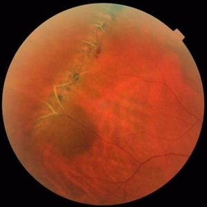
 Myopic CNV
Jan 11 2013 by Alex P. Hunyor, MD
Myopic macular degeneration complicated by subretinal neovascularisation, left eye.
Condition/keywords: high myopia, myopia, myopic choroidal neovascularization (CNV)
-
 RPE Reticular Degeneration
Jun 4 2014 by Henry J. Kaplan, MD
RPE reticular degeneration. #2
Condition/keywords: senile reticular degeneration
-
 Retinal Angiomatous Proliferation in Age-Related Macular Degeneration with Subretinal Neovascularization
Sep 24 2012 by James B. Soque, CRA, OCT-C, COA, FOPS
75-year-old white male with classic SRN with RAP. Lesion OD is active, and patient is receiving anti-VEGF treatment. Mid phase FA, 50 Deg, Mag 2x.
Photographer: James Soque, CRA, COA, Island Retina, Shirley, NY, USA
Imaging device: Topcon TRC 50 DX, OIS 5.0 MP Color, FA Camera, OIS Software
Condition/keywords: age-related macular degeneration (AMD), fundus autofluorescence (FAF), leakage, retinal angiomatous proliferation (RAP), subretinal neovascularization (SRNV)
-
 Lattice Degeneration and Choroidal Nevus
Oct 10 2015 by Hamid Ahmadieh, MD
Color fundus photograph of the right eye of a 46-year-old woman with a typical lattice degeneration and an adjacent choroidal nevus.
Photographer: Solmaz Shahmohammad, Negah Eye Center, Tehran, Iran
Condition/keywords: choroidal nevus, color fundus photograph, lattice degeneration
-
 Myopic Degeneration - Fundus Image
Oct 3 2013 by Gerardo Garcia-Aguirre, MD
Myopic degeneration - fundus image.
Condition/keywords: fundus photograph, myopic degeneration
-
 Pigmented Peripheral Retinal Degeneration
Jun 27 2013 by Jason S. Calhoun
42-year-old male came in for routine eye exam and to follow up on peripheral retinal degeneration in both eyes. VA is 20/20, right eye and 20/25, left eye. Patient is asymptomatic with no visual complaints.
Photographer: Jason S. Calhoun, Mayo Clinic Jacksonville, Florida
Imaging device: TOPCON TRC 50-EX
Condition/keywords: peripheral retinal degeneration
-
 Retinal Detachment with Macula Partially Detached
Oct 12 2012 by Jeffrey G. Gross, MD, FASRS
RD with macula partially detached.
Condition/keywords: macula, retinal degeneration
-
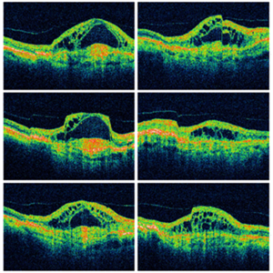
 Fibrovascular PED
Feb 21 2014 by Roy Schwartz, MD
72-year-old female with fibrovascular PED. Upper picture - PED with sub RPE hyper-reflective substance, in a multi-layered pattern, corresponding to fibrovascular PED. CME. Lower picture - PED flattened, a denser sub RPE hyperreflective substance is seen. CME resolved.
Condition/keywords: fibrovascular pigment epithelial detachment (PED), neovascular age-related macular degeneration (AMD), optical coherence tomography (OCT), ranibizumab
-
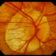 Peripapillary Atrophy With High Myopia
Feb 4 2015 by H. Michael Lambert, MD
Peripapillary atrophy and central macular degeneration seen in high myopia.
Condition/keywords: high myopia, peripapillary atrophy
-
 Retinal Break at Site of Lattice Degeneration with Scleral Indentation
Nov 9 2012 by Norman Byer
This is the same case as the previous photograph. With scleral indentation slightly more posterior, the flap is seen to be associated with a large retinal tear. This is a tractional tear and it is possible that in this case the cryotherapy itself may have increased the vitreoretinal traction at this site and in this way led to this new tear. The age of the tear is unknown because it was asymptomatic, and even though the eye is aphakic the tear has not caused a clinical retinal detachment.
Condition/keywords: retinal flap, scleral indentation, tractional retinal tear, vitreoretinal traction
 Loading…