Search results (1109 results)
-
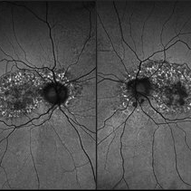
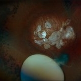
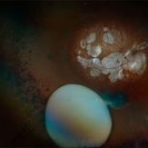
 Optic Disc Drusen
Aug 20 2025 by Drew Mitchell
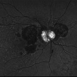
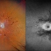
Fundus Autofluorescence photo of an 86 year old woman with neovascular AMD with active CNV and optic disc drusen.
Photographer: Drew Mitchell OCT-C
Imaging device: Optos California
Condition/keywords: fundus autofluorescence (FAF), neovascular age-related macular degeneration (AMD), optic disc drusen, OPTOS
-
 Subretinal Neovascular Membrane
Aug 15 2025 by Akansha Sharma
Color fundus photograph of a 40 year old male with subretinal neovascular membrane.
Photographer: DR. AKANSHA SHARMA
Condition/keywords: choroidal neovascular membrane (CNVM), CNVM, SRNVM, subretinal neovascularization (SRNV), wet age-related macular degeneration (wet AMD)
-
 Subretinal Neovascular Membrane
Aug 15 2025 by Akansha Sharma
Color fundus photograph of a 40 year old male with subretinal neovascular membrane.
Photographer: DR. AKANSHA SHARMA
Condition/keywords: choroidal neovascular membrane (CNVM), CNVM, Myopic CNVM, SRNVM, subretinal neovascularization (SRNV), Wet age related macular degeneration
-
 Retinal Detachment with Multiple Breaks
Aug 12 2025 by Kimberly Wakester
Optomap RGB of a 59-year-old man with a retinal detachment with multiple breaks in the left eye. Surgery was recommended. Patient is to continue follow up care post operatively.
Photographer: Kimberly Wakester, COA, OCT-C, Retina Consultants of Carolina
Imaging device: Optos California
Condition/keywords: lattice degeneration, left eye, Retinal Detachment with Multiple Breaks
-
 Secondary Pigmentary Degeneration of Retina
Jul 18 2025 by Kimberly Wakester
Optomap RGB and AF of an 63-year-old man with secondary pigmentary degeneration of the retina. Patient's Spark genetic testing revealed heterozygous mutations of unknown significance in LRP5, COL18A1, CPLANE1, SLC24A1 and VCAN. Clinical findings most consistent with Wagner's Syndrome (VCAN mutation, autosomal dominant). Will continue follow up care every 6 months with dilated exam and repeat OCT and Optos imaging.
Photographer: Kimberly Wakester, COA, OCT-C
Imaging device: Optos California
Condition/keywords: secondary pigmentary degeneration, Wagner's Syndrome
-
 Secondary Pigmentary Degeneration of Retina
Jul 18 2025 by Kimberly Wakester
Optomap RGB and AF of an 63-year-old man with secondary pigmentary degeneration of the retina. Patient's Spark genetic testing revealed heterozygous mutations of unknown significance in LRP5, COL18A1, CPLANE1, SLC24A1 and VCAN. Clinical findings most consistent with Wagner's Syndrome (VCAN mutation, autosomal dominant). Will continue follow up care every 6 months with dilated exam and repeat OCT and Optos imaging .
Photographer: Kimberly Wakester, COA, OCT-C
Imaging device: Optos California
Condition/keywords: secondary pigmentary degeneration, Wagner disease
-
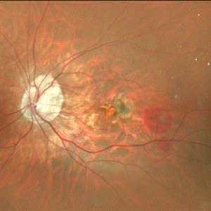
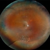
 Neovascular AMD with Active CNV
May 22 2025 by Kimberly Wakester
Optomap RGB of an 82-year-old man with Neovascular AMD with Active CNV and Dry AMD in the right eye. There is advanced atrophic changes without subfoveal involvement located temporally to the fovea. Patient is to continue follow up care with dilated exam, repeat OCT, and treatment of intravitreal injection of Vabysmo every 5 weeks at this time.
Photographer: Kimberly Wakester, COA, OCT-C
Imaging device: Optos California
Condition/keywords: advanced geographic atrophy, dry age-related macular degeneration (dry AMD), neovascular age-related macular degeneration (AMD)
-
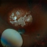
 Retromode Image of Dry AMD
May 13 2025 by Anupama Kiran Kumar
A retromode pseudo3D image of a case of dry AMD(NIDEK Mirante ). Retromode deviated left depicts depressed (pseudo concave) medium to large drusen. Here the drusenoid deposits appear to have a spectacular "moon surface" like appearance.
Photographer: Mr Pratap
Imaging device: Mirante SLO/OCT (Nidek Co., Gamagori, Japan)
Condition/keywords: AMD, dry age-related macular degeneration (dry AMD), retro mode
-
 Toxic Maculopathy (Elmiron)
Apr 9 2025 by Virginia Gebhart
79 year old male with toxic maculopathy from long term use of Elmiron (15+ yrs.) On exam there is stippled RPE changes, pigment clumping, and subretinal deposits. BCVA 20/100 | 20/40.
Photographer: Virginia Gebhart, Retina Consultants of Carolina
Imaging device: Optos California
Condition/keywords: autofluorescence imaging, cystoid macular degeneration, Elmiron Toxicity, Toxic Maculopathy
-
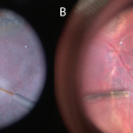
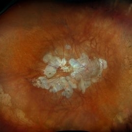
 Large Horseshoe Tear
Apr 4 2025 by Tejaswita Verma
Fundus photo of a 29 year old myopic male with RE 6/12P vision with large ragged tear on the posterior edge of lattice degeneration
Photographer: DR. TEJASWITA VERMA
Imaging device: MIRANTE
Condition/keywords: horseshoe tear
-
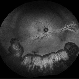
 Retinal Detachment and Lattice Degeneration
Mar 25 2025 by Korey Starkey
26 year-old patient presented at first visit with rhegmatogenous macula involving retinal detachment of the left eye. Underwent prompt surgical repair. Both eyes also present with lattice degeneration with atrophic holes.
Photographer: Korey Starkey
Condition/keywords: atrophic retinal hole, fundus photography, lattice degeneration, montage photo, Optos, OPTOS CALIFORNIA RGB, retinal detachment, retinal holes, rhegmatogenous retinal detachment, ultra-wide field imaging
-
 Cannula Tip Pressure
Mar 25 2025 by Robert Andrew Sisk, MD, FACS, FASRS
Color stills from surgical videos of subretinal delivery of gene augmentation therapy with A) voretigene neparvovec-ryzl and B) laru-zova. In the left panel, the cannula is slightly bent, and the retina and RPE are blanched white around the cannula tip engagement. The bleb was challenging to form in this patient with advanced retinal degeneration, and the bleb is shallow and mostly clear. In the right panel, the cannula tip is gently engaged, the cannula is straight, and it follows the retinotomy as the retina is elevated by the injection fluid.
Imaging device: Leica Proveo 8
Condition/keywords: gene therapy, genetic disorder, Leber's congenital amaurosis, retinitis pigmentosa, subretinal injection
-
 Lattice Degeneration With Atrophic Retinal Holes
Jan 30 2025 by Kimberly Wakester
Ultra-wide field montage fundus photograph of a 56-year-old woman with lattice degeneration with atrophic holes statues post laser. Patient also has a small CHRPE temporal to macula and trace ERM that is not visually significant. Will continue follow up care to monitor and treat as needed.
Photographer: Kimberly Wakester, COA
Imaging device: Optos California
Condition/keywords: atrophic retinal hole, CHRPE, epiretinal membrane (ERM), lattice degeneration, montage photo
-
 Elmiron Toxicity
Jan 15 2025 by Virginia Gebhart
54 year old female with pigmentary degeneration secondary to Elmiron. Stippled RPE maculopathy has lightly progressed with stable vision compared to previous visits. BCVA 20/200 OU. Pt reports taking Elmiron from 2010 to 2019.
Photographer: Virginia Gebhart
Imaging device: Optos California
Condition/keywords: autofluorescence imaging, Maculopathy, secondary pigmentary degeneration
-
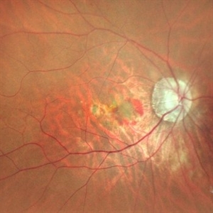
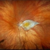 Subretinal Fibrosis
Jan 14 2025 by Kimberly Wakester
Fundus photograph of an 86-year-old woman with the end stage of Age-related Macular Degeneration in the left eye. Patient went unseen for 3-4 years prior to establishing care at our practice. Due to the significant amount of subretinal fibrosis, treatment was not recommended due to limited visual recovery. Patient was advised of monocular vision and the importance of follow up care.
Photographer: Kimberly Wakester, COA
Imaging device: Optos California
Condition/keywords: AMD, subretinal fibrosis
-
 New Subretinal Hemorrhage in AMD
Jan 8 2025 by Drew Mitchell
HD 1 line 100x OCT scan of a New Subretinal Hemorrhage in a established patient with AMD.
Photographer: Drew Mitchell, OCT-C
Imaging device: Zeiss Cirrus 6000
Condition/keywords: age-related macular degeneration (AMD), OCT, subretinal hemorrhage
-
OCT Video Imaging of Left Eye Age Related Macular Degeneration
Jan 6 2025 by Kavitha Duraipandi, MD DNB FICO FRCS
Left eye OCT macula shows various biomarkers like PED, sub retinal fluid, sub retinal hyper reflective material and hyper reflective foci suggestive of wet age-related macular degeneration.
Condition/keywords: OCT biomarkers, wet age-related macular degeneration (wet AMD)
-
 Subluxation of the Lens
Dec 12 2024 by Kimberly Wakester
Ultra-wide field fundus photos of an 53-year-old man with a Subluxation of the Lens in the posterior vitreous cavity of the right eye after a trauma that happened many years ago. Patient remains stable with no adverse reaction to the lens at this time. No surgical intervention is recommended at this time. Patient also has myopic degeneration and lattice degeneration that will require patient to have follow up care.
Photographer: Kimberly Wakester, COA
Imaging device: Optos California
Condition/keywords: lattice degeneration, myopic degeneration, peripapillary atrophy, posterior staphyloma, Subluxation of the Lens
-
 Myopic Degeneration
Dec 9 2024 by Virginia Gebhart
67 year old female with myopic degeneration. Posterior staphylomas are stable. VA limited by extensive chorioretinal atrophy. BCVA 20/160 (ecc)
Photographer: Virginia Gebhart, Retina Consultants of Carolina
Imaging device: Optos California
Condition/keywords: chorioretinal atrophy, myopic degeneration, staphyloma
-
 Repaired Retinal Detachment with Multiple Breaks
Dec 9 2024 by Virginia Gebhart
FAF in 25 year old female of repaired retinal detachment 1.5 year s/p scleral buckle/cryo. Pt had been having symptoms for over a year, inferior demarcation line from retinal fluid that was present. Retina remains flat and attached under buckle. Treated lattice inferiorly, no new holes or tears. VA 20/20
Photographer: Virginia Gebhart, Retina Consultants of Carolina
Imaging device: Optos California
Condition/keywords: autofluorescence imaging, cryotherapy, demarcation line, lattice degeneration, scleral buckle
-
 Macular Degeneration
Dec 3 2024 by Sarah D Kang
Fundus photograph of an 85-year-old female patient with macular degeneration observed for retinal clearance before cataract surgery.
Condition/keywords: floaters, macular degeneration
-
 Macular Degeneration
Dec 3 2024 by Sarah D Kang
Fundus photograph of an 85-year-old female patient with macular degeneration observed for retinal clearance before cataract surgery.
Condition/keywords: floaters, macular degeneration
-
 Pigmentary Degeneration of Retina (Secondary to Elmiron)
Nov 27 2024 by Virginia Gebhart
77 year old female with advanced geographic atrophy after years of Elmiron use (stopped in 2018). Serial exams show continued progression of GA. Central vision limited, vision remains stable and patient does not report noticing any changes.
Photographer: Virginia Gebhart, Retina Consultants of Carolina
Imaging device: Optos California
Condition/keywords: geographic atrophy, secondary pigmentary degeneration, toxic maculopathy
 Loading…