Search results (1109 results)
-
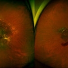
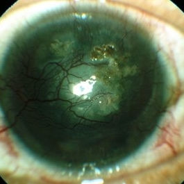 Corneal Vascularisation With Spheroidal Degeneration
Jun 3 2017 by S. Natarajan, MD, FASRS, FRCS (GLASGOW) , FICO, D.Sc, FELA
Fundus photograph of a 50-year-old male corneal vascularization with spheroidal degeneration.
Photographer: miss ashwini borde
Imaging device: call zeiss 450 plus IR
Condition/keywords: corneal vascularization, degeneration
-
 Keratoconus
Oct 2 2013 by Jerald A. Bovino, MD
This is a profile picture of a patient with keratoconus.
Condition/keywords: cornea, degeneration, ectasia, thinning
-
 Ocular Albinism Carrier
Feb 13 2013 by From the Collections of Thomas M. Aaberg, MD and Thomas M. Aaberg Jr., MD
RPE atrophy, degeneration, thinned arterioles.
Condition/keywords: degeneration, ocular albinism, retinal pigment epithelium atrophy, thinned arterioles
-
 Ocular Albinism Carrier
Feb 13 2013 by From the Collections of Thomas M. Aaberg, MD and Thomas M. Aaberg Jr., MD
RPE atrophy, degeneration, thinned arterioles.
Condition/keywords: degeneration, ocular albinism, retinal pigment epithelium atrophy, thinned arterioles
-
 Slide 4-5
Feb 20 2019 by Lancaster Course in Ophthalmology
Retinitis pigmentosa (primary pigmentary degeneration), an example of a disease with a recessive pattern of inheritance. Pigment clumping in characteristic bone-corpuscle pattern.
Condition/keywords: degeneration, retinitis pigmentosa
-
 Slide 4-7
Feb 20 2019 by Lancaster Course in Ophthalmology
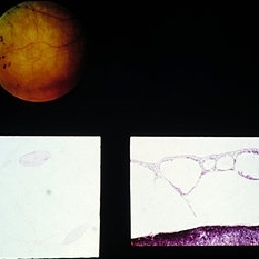
Primary pigmentary degeneration of the retina. Histologic section shows clumps of pigment around the , and atrophy of all retinal layers, including the pigment epithelium ( x40). {Massachusetts Eye and Ear Infirmary, No. E49-274.)
Condition/keywords: atrophy, degeneration, pigment epithelium, retinal capillaritis
-
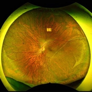
 Slide 9-55
Feb 26 2019 by Lancaster Course in Ophthalmology
Cystic degeneration of retina overlying a small choroidal malignant melanoma. Fluorescein staining of the cystic spaces is present late in transit.
Condition/keywords: degeneration, melanoma
-
 Degeneration Paravenous
Sep 20 2016 by JEFFERSON R SOUSA, Tecg.º (Biomedical Systems Technology)
Female patient, 32-years-old, Asian, appeared at the clinic with a history of glaucoma. 20/20 visual acuity in both eyes. Examination of color photography, pigmentary changes were observed following the vascular arcades only in the left eye. Suggestive of paravenous degeneration.
Photographer: JEFFERSON R SOUSA - Study Center and Ophthalmological Research Dr. Andre M V Gomes, Institute Dr. Suel Abujamra São Paulo-Brazil
Imaging device: Zeiss / VisuCam-500 - Angulation of field photo of 45 Degrees, flash 11.
Condition/keywords: degeneration paravenous
-
 Degeneration Paravenous
Sep 20 2016 by JEFFERSON R SOUSA, Tecg.º (Biomedical Systems Technology)
Female patient, 32-years-old, Asian, appeared at the clinic with a history of glaucoma. 20/20 visual acuity in both eyes. Examination of color photography, pigmentary changes were observed following the vascular arcades only in the left eye. Suggestive of paravenous degeneration.
Photographer: JEFFERSON R SOUSA - Study Center and Ophthalmological Research Dr. Andre M V Gomes, Institute Dr. Suel Abujamra São Paulo-Brazil
Imaging device: Zeiss / VisuCam-500 - Angulation of field photo of 45 Degrees, flash 17.
Condition/keywords: degeneration paravenous
-
 Degeneration Paravenous
Sep 20 2016 by JEFFERSON R SOUSA, Tecg.º (Biomedical Systems Technology)
Female patient, 32-years-old, Asian, appeared at the clinic with a history of glaucoma. 20/20 visual acuity in both eyes. Examination of color photography, pigmentary changes were observed following the vascular arcades only in the left eye. Suggestive of paravenous degeneration.
Photographer: JEFFERSON R SOUSA - Study Center and Ophthalmological Research Dr. Andre M V Gomes, Institute Dr. Suel Abujamra São Paulo-Brazil
Imaging device: Zeiss / VisuCam-500 - Angulation of field photo of 45 Degrees, flash 24.
Condition/keywords: degeneration paravenous
-
 Degeneration Paravenous
Sep 20 2016 by JEFFERSON R SOUSA, Tecg.º (Biomedical Systems Technology)
Female patient, 32-years-old, Asian, appeared at the clinic with a history of glaucoma. 20/20 Visual acuity in both eyes. Examination of color photography, pigmentary changes were observed following the vascular arcades only in the left eye. Suggestive of paravenous degeneration.
Photographer: JEFFERSON R SOUSA - Study Center and Ophthalmological Research Dr. Andre M V Gomes, Institute Dr. Suel Abujamra São Paulo-Brazil
Imaging device: Zeiss / VisuCam-500 - Angulation of field photo of 45 Degrees, flash 11.
Condition/keywords: degeneration paravenous
-
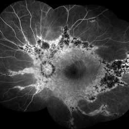
---thumb.JPG/image-square;max$300,300.ImageHandler) "Flower" Macular Degeneration (Wet)
"Flower" Macular Degeneration (Wet)
Jul 13 2013 by Jason S. Calhoun
Patient with (wet) macular degeneration in the left eye. Notice the "flower" shape abnormal blood vessels staining.
Photographer: Jason S. Calhoun, Department of Ophthalmology, Mayo Clinic Jacksonville, Florida
Imaging device: TOPCON TRC 50-EX
Condition/keywords: choroidal neovascularization (CNV)
-
 1 year Follow Up after Scleral Buckle Surgery in a Young Patient
May 18 2023 by Jesus Lozano, MD
25 year old man after Scleral Buckle Surgery + laser Retinopexy do to RRD macula off with ínfero temporal mid peripheral retinal holes in an area of lattice degeneration. Final VA 6/9.
Imaging device: Optos
Condition/keywords: scleral buckle
-
---thumb.jpg/image-square;max$300,300.ImageHandler) Abnormal Fundus
Abnormal Fundus
Oct 15 2013 by Maurice F. Rabb
The patient is a 48 year old female who was noted to have an abnormal fundus on routine examination. Her past medical history is unremarkable. Her past family history is remarkable for her father being diagnosed with macular degeneration at age 72. Visual acuity was 20/20-1, J-2, OD and 20/20, J-1, OS. Amsler grid examinaton was normal, OU. All 6 of the AOHRR screening plates were identified correctly, OU. The 45 minute rod psychophysiologic threshold was normal, OU. ERG responses were normal for both rod and cone amplitudes. The cone implicit times were normal. The EOG was normal.
Condition/keywords: abnormal fundus
-
 Active Neovascular AMD With Disciform Scar
Apr 30 2015 by Mitzy E Torres Soriano, MD
Active neovascular AMD with disciform scar.
Photographer: Mitzy E. Torres Soriano, MD; Centro medico Cagua-Estado Aragua. Venezuela
Imaging device: TOPCON
Condition/keywords: disciform scar, disciform with hemorrhage, neovascular age-related macular degeneration (AMD), wet age-related macular degeneration (wet AMD)
-
 Acute Posterior Vitreous Detachment
Nov 9 2012 by Norman Byer
This large and complicated retinal tear in a 51-year-old man resulted from an acute posterior vitreous detachment which concentrated its tractional forces around this area of lattice degeneration. Because of the powerful traction, there is an additional central tear splitting the large retinal flap and almost severing one of its arms. The traction was strong enough to completely rupture the blood vessel just to the left of the flap. Marking the ruptured peripheral end of the blood vessel is a yellow depigmented thrombus.
Condition/keywords: acute posterior vitreous detachment, depigmented thrombus, lattice degeneration, retinal tear, tractional retinal detachment
-
 Acute Retinal Detachment
Nov 9 2012 by Norman Byer
This 54-year-old man was referred because of sudden symptoms in his opposite eye in which he had suffered an acute retinal detachment secondary to a horseshoe tear around lattice degeneration. During the examination, the fellow eye shown here was also found to have this large horseshoe tear about 1 o’clock hour (4 disc diameters) in size. A tear occurred around a lattice lesion which is present on the flap but is out of focus. This tear had been asymptomatic even though it was caused by a posterior vitreous detachment and illustrates that even very large tears may produce no symptoms or mild symptoms that are easily overlooked.
Condition/keywords: lattice degeneration, posterior vitreous detachment
-
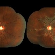
 Advanced Angioid Streak-Associated Choroidal Neovasclar Membranes
Dec 27 2016 by Young Hee Yoon, MD, PhD
UWF fundus photographs of an 74-year-old woman who received several anti-VEGF injections due to CNV associated with angioid streak in both eyes. Note diffuse scar change and pigmentary degenerations aroud disc in both eyes. There is some retinal hemorrhage due to CNV in her left eye.
Photographer: Young Hee Yoon, University of Ulsan, Asan Medical Center, Seoul, Korea
Imaging device: Optomap
Condition/keywords: angioid streaks, choroidal neovascularization (CNV)
 Loading…