Search results (1109 results)
-
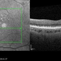
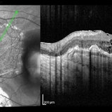
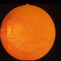
 Cuticular and soft drusen
Jun 14 2021 by Gerardo Garcia-Aguirre, MD
Fundus photograph (left) and Retro mode infrared image (right) of an eye with soft and cuticular drusen. Drusen are highlighted and better visualized with retro mode imaging.
Photographer: Gerardo Garcia-Aguirre
Imaging device: Nidek Mirante
Condition/keywords: drusen, dry age-related macular degeneration (dry AMD)
-
 Lady in a dress
Feb 9 2023 by Shelby Helton
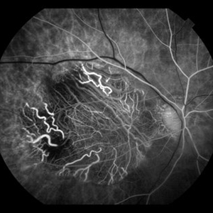
Fluorescein Angiography on a 67-year-old male with significant RPE changes secondary to a severe subretinal hemorrhage that required a vitrectomy with subretinal TPA in 2013.
Photographer: Shelby Helton
Imaging device: Heidelberg Spectralis
Condition/keywords: wet age-related macular degeneration (wet AMD)
-
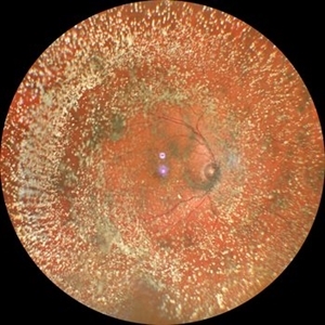
 Rod Cone dystrophy
Nov 29 2022 by Niloofar Piri, MD
Fundus autofluorescence of the left eye in a 58 yo male with rod cone dystrophy. He presented with night blindness and peripheral vision loss since youth and recent decrease in central vision for the past 10 years. Notice multiple coin shaped hypoautofluorescent pacthes within central 20 degrees which are coalescing centrally. (fundus photo uploaded separately) He has one pathogenic variants of both CEP290 and PRPH2 genes.
Photographer: Sean Kelso, Saint Louis University
Condition/keywords: hereditary retinal degeneration, hereditary retinal dystrophy, rod cone dystrophy
-
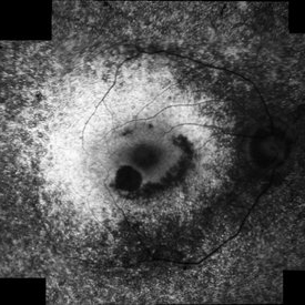
 Cuticular Drusen
Jan 17 2024 by John Lee
Heidelberg SD-OCT of a 65-year-old woman with age-related macular degeneration demonstrating classic sawtooth appearance of cuticular drusen.
Photographer: Natasha Vinson
Imaging device: Heidelberg Spectralis
Condition/keywords: age-related macular degeneration (AMD), cuticular drusen
-
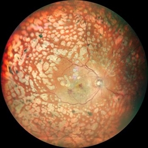
 Diffuse Chorioretinal Atrophy
Feb 21 2024 by Virginia Gebhart
61 year male with myopic degeneration and diffuse chorioretinal atrophy. BCVA 20/200.
Photographer: Virginia Gebhart
Imaging device: Topcon TRC 50DX
Condition/keywords: chorioretinal atrophy, myopic degeneration
-
 Dry AMD
Jun 4 2014 by Henry J. Kaplan, MD
Multiple drusen with RPE changes in the macula #2.
Condition/keywords: age-related macular degeneration (AMD), dry age-related macular degeneration (dry AMD)
-
 Dry AMD, Advanced Atrophic without Subfoveal Involvement
Oct 12 2021 by Kelli Nyenhuis
OCT Heidelberg photograph of a 79-year-old woman with AMD advancing that has been observed over the last 10 years.
Photographer: Kelli Nyenhuis, OMA
Imaging device: Heidelberg
Condition/keywords: dry age-related macular degeneration (dry AMD)
-
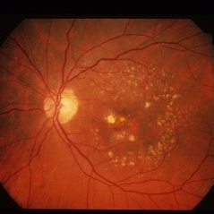
 Geographic Atrophy
Oct 13 2012 by Geoffrey G. Emerson, MD, PhD, FASRS
Geographic atrophy
Condition/keywords: advanced geographic atrophy, choroid, dry age-related macular degeneration (dry AMD)
-
 Massive SRH in Patient on Coumadin Being Treated for Exudative AMD
Sep 30 2019 by John S. King, MD
78-year-old white female using 1mg of warfarin for atrial fibrillation, who had a large PED, Type 1 lesion from AMD. Noticed acute darkening of vision one week after anti-VEGF injection. Has very large SRH, subRPE heme, and corrugated retinal appearance post RPE tear. Vision HM (from 20/100). 20/25 in the fellow eye that has dry AMD.
Photographer: Shelly Blair
Imaging device: Optos CA
Condition/keywords: subretinal hemorrhage, wet age-related macular degeneration (wet AMD)
-
 Normal Temporal Ora Serrata
Nov 9 2012 by Norman Byer
This is the normal temporal ora serrata in a 26-year-old man. Note the typical ragged moth-eaten appearance caused by peripheral cystoid degeneration. This appearance may be present in infants but is always present beyond the age of eight years.
Condition/keywords: ora serrata, peripheral cystoid degeneration
-
 Prominent Long Ciliary Nerve
Jan 25 2022 by Kachelle Brown
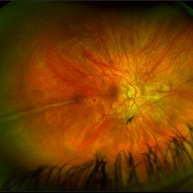
Ultra-wide field photograph of a 48-year-old female with a prominent long ciliary nerve. Patient presented asymptomatic, and was referred for a macula on retinal detachment. Patient was diagnosed with high myopia and a posterior vitreous detachment, and the physician discussed increased risk of floaters, myopic degeneration and retinal detachment associated with high myopia. -24.00 prior to cataract surgery OU per patient.
Photographer: Kachelle Brown
Imaging device: Optos California
Condition/keywords: fundus photograph, high myopia, long ciliary nerve, optos, right eye, ultra-widefield image
-
 Retinal Tear at the Posterior Edge of Lattice Degeneration
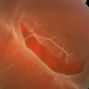
Mar 1 2014 by Homayoun Tabandeh, MD, FASRS
Retinal tear at the posterior edge of lattice degeneration.
Condition/keywords: lattice degeneration, retinal tear
-
 RPE-Transplantation
Jul 25 2024 by Gabriel Costa Andrade, PhD
Postoperative period of RPE-transplantation in a patient with neovascular AMD after RPE tear.
Photographer: Gabriel Andrade
Condition/keywords: neovascular age-related macular degeneration (AMD), pars plana vitrectomy (PPV), wet age-related macular degeneration (wet AMD)
-
 Snail Track Peripheral Retinal Degeneration
Apr 29 2022 by Otakar Dušek, M.D. Ph.D.
Colour fundus photograph of 22-year-old woman with incidentally found snail track retinal degeneration in the superior temporal periphery of the retina of the right eye.
Photographer: Otakar Dušek, Charles University, Prague
Imaging device: Zeiss Clarus
Condition/keywords: peripheral retinal degeneration
-
---thumb.jpg/image-square;max$300,300.ImageHandler) Age Related Macular Degeneration - Geographic Atrophy
Age Related Macular Degeneration - Geographic Atrophy
May 3 2013 by Suber S. Huang, MD, MBA, FASRS
Geographic Atrophy.
Imaging device: Retina Diseases Imaging Analysis Reading Center
Condition/keywords: advanced geographic atrophy, atrophic scar, atrophic spot, geographic atrophy, macula lesion, pigment epithelial atrophy
-
 AGE RELATED MACULAR DEGENERATION AUTOFLUORESCENCE
Aug 13 2023 by Aditya S Kelkar, MS, FRCS, FASRS,FRCOphth
Autofluorescence fundus photography of an 78-year-old woman diagnosed with age-related macular degeneration.
Photographer: Dr. Harsh Jain, National Institute of Ophthalmology
Imaging device: Clarus 500
Condition/keywords: age-related macular degeneration (AMD)
-
 AMD
AMD
Jul 26 2014 by Avris Romario Diparaja Siahaan
An autofluorescence image of a 78-year-old-man with an age-related macular degeneration on his both eyes.
Photographer: Avris Romario Diparaja Siahaan, Klinik Mata Nusantara
Imaging device: Heidelberg Spectralis
Condition/keywords: age-related macular degeneration (AMD), autofluorescence imaging
-
 Angioid Streaks
Jan 20 2021 by Nivesh Gupta
Fundus photograph of an 51-year-old female patient with angioid streaks with secondary choroidal neovascular membrane.
Photographer: Nivesh Gupta, Retina Fellow, Retina Foundation, Ahmedabad, India
Imaging device: NIDEK SLO MIRANTE
Condition/keywords: age-related macular degeneration (AMD), angioid streaks, choroidal neovascular membrane (CNVM)
-
 ARMD
ARMD
Jan 13 2014 by David Callanan, MD
HM OU marked RPE atrophy, 63-year-old female.
Condition/keywords: age-related macular degeneration (AMD)
-
 Asteroid hyalosis
Dec 1 2023 by Anjana Mirajkar, MS Ophthalmology
A widefield image of RE of a 65 year old male with dense asteroid Hyalosis
Photographer: Dr. Anjana Mirajkar -Retina Foundation, Ahmedabad
Imaging device: Mirante-Nidek
Condition/keywords: asteroid hyalosis, vitreous degeneration
-
 Cancer-Associated Retinopathy (CAR)
Jun 30 2018 by Peter G. Hovland, MD, PhD
Autofluorescence image of affected right eye of 59-year-old woman 6 years after onset of cancer-associated retinopathy. Demonstrates extensive RPE degeneration.
Photographer: Colorado Retina Associates
Imaging device: Heidelberg Spectralis
Condition/keywords: retinal pigment epithelium, retinopathy
-
 Choroideremia
Sep 21 2022 by Zach Seim
Ultra-widefield fundus photo of a 74 year old male presenting with severe vision loss beginning at age 55. Patient sought a second opinion with our office and was diagnosed with Choroideremia. Patient denies hearing loss, heart problems, balance issues, polydactyly, kidney problems, and dental problems. Patient reports that nobody in the family had blindness. Choroideremia is an X-linked chorioretinal dystrophy characterized by the diffuse, progressive degeneration of the retinal pigment epithelium (RPE), photoreceptors and choriocapillaris. It is caused by a mutation in the CHM gene.
Photographer: Zach Seim
Imaging device: Optos California
Condition/keywords: choroideremia, hereditary choroidal atrophy, hereditary retinal dystrophy, Optos, pseudocolor, ultra-wide field imaging
-
 Choroideremia
Sep 21 2022 by Zach Seim
Ultra-widefield fundus photo of a 74 year old male presenting with severe vision loss beginning at age 55. Patient sought a second opinion with our office and was diagnosed with Choroideremia. Patient denies hearing loss, heart problems, balance issues, polydactyly, kidney problems, and dental problems. Patient reports that nobody in the family had blindness. Choroideremia is an X-linked chorioretinal dystrophy characterized by the diffuse, progressive degeneration of the retinal pigment epithelium (RPE), photoreceptors and choriocapillaris. It is caused by a mutation in the CHM gene.
Photographer: Zach Seim
Imaging device: Optos California
Condition/keywords: choroideremia, hereditary choroidal atrophy, hereditary retinal dystrophy, left eye, light perception, low vision, Optos, pseudocolor, ultra-wide field imaging
-
 Chronical Submacular Hemorrhage in the Setting of Neovascular AMD
Mar 23 2015 by Rita Couceiro, MD, MS
An 80-year-old male, with a history of hypertension and high cholesterol, complained of acute and painless vision loss in his left eye (OS) in the previous 5 months. On observation best corrected visual acuity in OS was hand motion. A dense vitreous opacity in OS precluded fundus examination. Ocular ultrasound revealed vitreous hemorrhage and thickening of the macular area. The patient was submitted to pars plana vitrectomy, which disclosed a large submacular hemorrhage with chronical features and disciform scarring in the setting of neovascular AMD.
Imaging device: Intraoperative fundus photograph
Condition/keywords: neovascular age-related macular degeneration (AMD), submacular hemorrhage, wet age-related macular degeneration (wet AMD)
-
 CNVM in Pan-retinal Photocoagulated Patient
Dec 30 2020 by ASRS Staff
Wide fundus photograph of 65-year-old, female, diabetic patient.
Imaging device: Nidek Mirante
Condition/keywords: age-related macular degeneration (AMD), diabetes, pan-retinal photocoagulation (PRP)
 Loading…