Search results (34 results)
-
 Vitreous Hemorrhage
Jul 10 2018 by Karen Panzegrau
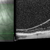
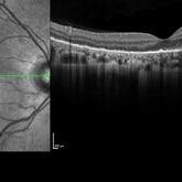
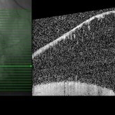
SD-OCT of a 35-year-old female presenting with a vitreous hemorrhage of her left eye. Patient has active proliferative diabetic retinopathy, as well as a completed posterior vitreous detachment in the left eye.
Photographer: Karen Panzegrau
Condition/keywords: diabetes, Heidelburg Spectralis, left eye, optical coherence tomography (OCT), posterior vitreous detachment, proliferative diabetic retinopathy (PDR), vitreous hemorrhage
-
 Neovascular AMD with Active CNV
Jan 2 2018 by Carolyn Daley
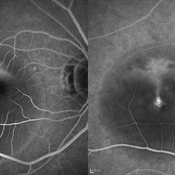
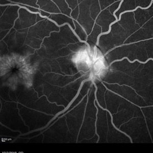
30 degree fluorescein angiogram of an 80-year-old woman with neovascular AMD with active CNV in the left eye. Patient is being treated with Avastin.
Photographer: Carolyn Daley, Retina Specialists of Michigan
Imaging device: Heidelberg Spectralis
Condition/keywords: 30 degrees, choroidal neovascularization (CNV), Heidelburg Spectralis, left eye, neovascular age-related macular degeneration (AMD)
-
 Coats Disease
May 27 2016 by Olivia Rainey
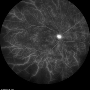
Composite fluorescein angiogram of the left eye of a man with Coats Disease.
Photographer: Olivia Rainey
Imaging device: Heidelberg Spectralis
Condition/keywords: Coats' disease, composite, fluorescein angiogram (FA), fluorescein leakage, Heidelburg Spectralis
-
 Macula Off Retinal Detachment
Jan 2 2018 by Carolyn Daley
55-year-old with macula off retinal detachment post cataract surgery.
Photographer: Carolyn Daley, Retina Specialists of Michigan
Imaging device: Heidelberg Spectralis
Condition/keywords: Heidelburg Spectralis, optical coherence tomography (OCT)
-
 Multiple Astrocytic Hamartomas
Jul 26 2018 by Olivia Rainey
Optical coherence tomography of a 7-year-old female with multiple astrocytic harmartomas as a retinal manifestation of tuberous sclerosis. Patient came to our office to rule out possible drug toxicity from Sabril, a an anticonvulsant. There were no signs of retinal toxicity by extended ophthalmoscopy or imaging, yet she will be monitored every 6 months.
Photographer: Olivia Rainey
Imaging device: Heidelberg Spectralis
Condition/keywords: astrocytic hamartoma, Heidelburg Spectralis, infrared image, left eye, optical coherence tomography (OCT), tuberous sclerosis
-
 Mild Nonproliferative Diabetic Retinopathy
Jan 16 2019 by Carolyn Daley
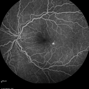
Fluorescence angiogram 50 degree imaging of a 38-year-old woman with mild nonproliferative diabetic retinopathy in the left eye. Patient also presented with a paracentral scotoma which etiologies could include vascular occlusion vs JFRT.
Photographer: Carolyn Daley, Retina Specialists of Michigan
Imaging device: Heidelberg Spectralis
Condition/keywords: diabetes, Heidelburg Spectralis, juxtafoveal telangiectasis, nonproliferative diabetic retinopathy, retinopathy, vascular occlusions
-
 Optic Nerve Head Drusen
Feb 9 2018 by Olivia Rainey
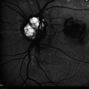
Fundus autofluorescence of a 49-year-old female with optic nerve head drusen affecting her left eye. The patient has pseudoxanthoma elasticum with choroidal neovascularization and has been receiving anti-VEGF treatment for many years.
Photographer: Olivia Rainey
Imaging device: Heidelberg Spectralis
Condition/keywords: 30 degrees, anti-VEGF, choroidal neovascularization (CNV), fundus autofluorescence (FAF), Heidelburg Spectralis, left eye, optic disc, optic nerve drusen, pseudoxanthoma elasticum (PXE)
-
 Posterior Uveitis with Cystoid Macular Edema
Jan 18 2018 by Olivia Rainey
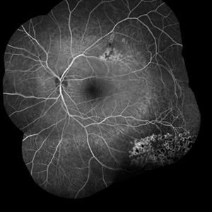
Ultra-wide field fluorescein angiogram of a 59-year-old female with posterior uveitis and chronic cystoid macular edema affecting her left eye. Interestingly, she has peripheral capillary nonperfusion inferotemporal, which could be driving CME.
Photographer: Olivia Rainey
Imaging device: Heidelberg Spectralis
Condition/keywords: 102 degrees, cystoid macular edema (CME), fluorescein leakage, Heidelburg Spectralis, left eye, peripheral retinal nonperfusion, posterior uveitis, ultra-wide field imaging
-
 Acute Macular Neuroretinopathy
Dec 11 2019 by Lauren Whaley
34-year-old female patient presented with changes in vision after recent upper respiratory infection. Referring doctor originally thought it was a blood pressure issue. She noticed a "C" shape in her vision. Infrared image was captured showing exactly what patient was describing! Doctor confirmed with this image that it was AMN.
Photographer: Lauren R. Whaley, COA
Imaging device: Heidelberg Spectralis
Condition/keywords: 30 degrees, acute macular neuroretinopathy, Heidelburg Spectralis, left eye, macula, near infrared autofluorescence (NIRAF)
-
 Plateau Iris from Aqueous Misdirection
Jan 18 2018 by Olivia Rainey
Anterior segment OCT of a 90-year-old female with plateau iris from aqueous misdirection affecting her right eye.
Photographer: Olivia Rainey
Imaging device: Heidelberg Spectralis
Condition/keywords: anterior segment, aqueous misdirection, Heidelburg Spectralis, optical coherence tomography (OCT), plateau iris
-
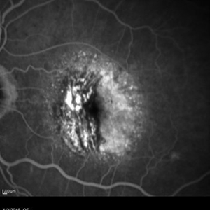
 Serpiginous Choroidal Atrophy
Mar 29 2019 by Jessica Norkus
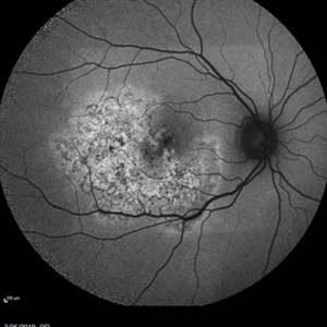
50 degree Auto fluorescent image of 20-year-old female presenting with serpiginous choroidal atrophy. Patient was unaware of vision loss OD, until accidentally covering OS and noticing the change. Acuity of 20/200 OD and 20/15 OS at time of imaging.
Photographer: Jessica Norkus
Imaging device: Heidelberg Spectralis
Condition/keywords: Heidelburg Spectralis, macula lesion, macula serpiginous choroidopathy, optical coherence tomography (OCT), wide angle imaging
-
 Central Serous Retinopathy
May 16 2017 by Olivia Rainey
Simultaneous fluorescein and indocyanine green angiography of an 37-year-old male with central serous retinopathy affecting his right eye. Patient's vision declined from 20/25 to 20/80 in the right eye. He elected for treatment with photodynamic therapy.
Photographer: Olivia Rainey
Imaging device: Heidelberg Spectralis
Condition/keywords: 30 degrees, central serous retinopathy (CSR), fluorescein angiogram (FA), fluorescein leakage, Heidelburg Spectralis, indocyanine green (ICG) angiography, late phase, mushroom cloud
-
 Peripheral Retinoschisis
Jul 26 2018 by Olivia Rainey
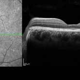
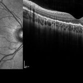
Optical coherence tomography of a 49-year-old male with non-progressive peripheral retinoschisis of his left eye. Patient was asymptomatic and had no prior trauma or surgery to his eye. Recommended observation at this time.
Photographer: Olivia Rainey
Imaging device: Heidelberg Spectralis
Condition/keywords: Heidelburg Spectralis, left eye, optical coherence tomography (OCT), retinoschisis
-
 Cystoid Macular Edema Secondary to Panuveitis
Jan 15 2019 by Olivia Rainey
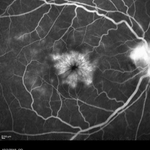
Fluorescein angiogram of a 55-year-old female with cystoid macular edema secondary to uveitis affecting her right eye. Patient was diagnosed with sarcoidosis.
Photographer: Olivia Rainey
Imaging device: Heidelberg Spectralis
Condition/keywords: 30 degrees, cystoid macular edema (CME), fluorescein angiogram (FA), fluorescein leakage, Heidelburg Spectralis, sarcoidosis, uveitis
-
 Plateau Fovea with Inner Retinal Thinning
May 27 2020 by Olivia Rainey
Optical coherence tomography of the left eye of a 20-year-old male with Alport Syndrome. The patient did not present with any ocular or visual symptoms, yet the distinct "plateau contour" of his fovea was noted on OCT during his visit. The patient presented with 20/25 vision at the time of his visit. There was myelinated nerve fiber layer noted in both eyes, but these features had remained stable from his appointment three years prior. The physician noted that myelinated nerve fiber was a congenital change, and had not affected his vision or health of the eye, nor is a feature of Alport Syndrome.
Photographer: Olivia Rainey, OCT-C, COA
Imaging device: Heidelberg Spectralis
Condition/keywords: Alports disease, Heidelburg Spectralis, inner retinal thinning, left eye, optical coherence tomography (OCT), plateau fovea
-
 Weiss Ring
Jan 15 2019 by Olivia Rainey
Fluorescein angiogram of a 55-year-old female with a Weiss ring affecting her right eye. Patient was diagnosed with sarcoidosis. She has cystoid macular edema secondary to panuveitis.
Photographer: Olivia Rainey
Imaging device: Heidelberg Spectralis
Condition/keywords: 30 degrees, cystoid macular edema (CME), fluorescein angiogram (FA), fluorescein leakage, Heidelburg Spectralis, optic nerve, sarcoidosis, uveitis, Weiss ring
-
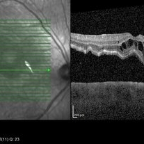
 Serpiginous Choroidal Atrophy
Mar 29 2019 by Jessica Norkus
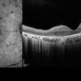
Heidelberg single horizontal scan image of 20-year-old female presenting with serpiginous choroidal atrophy. Patient was unaware of vision loss OD, until accidentally covering OS and noticing the change. Acuity of 20/200 OD and 20/15 OS at time of imaging.
Photographer: Jessica Norkus
Imaging device: Heidelberg Spectralis
Condition/keywords: Heidelburg Spectralis, macula lesion, macula serpiginous choroidopathy, optical coherence tomography (OCT)
-
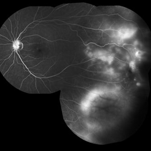
 Proliferative Diabetic Retinopathy
Jan 18 2018 by Olivia Rainey
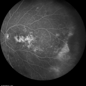
Ultra-wide field fluorescein angiogram of a 57-year-old male with proliferative diabetic retinopathy affecting his right eye.
Photographer: Olivia Rainey
Imaging device: Heidelberg Spectralis
Condition/keywords: diabetes, fluorescein leakage, Heidelburg Spectralis, neovascularization of the disc (NVD), peripheral retinal nonperfusion, proliferative diabetic retinopathy (PDR), ultra-wide field imaging
-
 Coats' Disease
Aug 24 2018 by Kim Barrett
Montage fluorescein angiography of 14-year-old male with Coats' Disease of the left eye. Multiple focal laser treatments. Current uncorrected visual acuity is 20/15-1 OU.
Photographer: Kim Barrett, C.O.A. Retina Specialist of Michigan
Imaging device: Heidelberg Spectralis
Condition/keywords: adolescent, Coats' disease, fluorescein angiogram (FA), Heidelburg Spectralis, laser photocoagulation, left eye, macroaneurysm, montage
-
 Multicolor Imaging in Diabetic Retinopathy
Sep 25 2018 by samarth mishra
A 60-year-old male presented with a history of blurring of vision since many months. He had a history of diabetes since last 8 years. On routine examination proliferative diabetic retinopathy with diabetic macular edema was noted. Fundus fluorescein angiography showed neovascularization elsewhere. Hard exudates can be seen as greenish yellow dots all over the posterior pole in multicolor imaging. Retinal hemorrhage can be seen as dark red.
Photographer: Aditya Birla Sankara Nethralaya, Kolkata, West Bengal , India
Condition/keywords: diabetic retinopathy, Heidelburg Spectralis, multicolor, optical coherence tomography (OCT)
-
 Multicolor Imaging of Bilateral Branch Retinal Vein Occlusion
Sep 25 2018 by samarth mishra
A 40-year-old female presented with complains of blurring of vision since past 1 week. Patient had a history of hypertension. On routine examination bilateral branch retinal vein occlusion was noted. Visual acuity at presentation was 6/9 and 6/15 in the right and left eye respectively. Multicolor composite imaging shows the hemorrhage as red and the retinal thickening as greenish hue. She was managed with anti vascular endothelial growth factor in both eyes.
Photographer: Aditya Birla Sankara Nethralaya, Kolkata, West Bengal , India
Condition/keywords: branch retinal vein occlusion (BRVO), Heidelburg Spectralis, multicolor, optical coherence tomography (OCT)
-
 Peripheral Retinoschisis
Jul 26 2018 by Olivia Rainey
Optical coherence tomography of a 49-year-old male with non-progressive peripheral retinoschisis of his left eye. Patient was asymptomatic and had no prior trauma or surgery to his eye. Recommended observation at this time.
Photographer: Olivia Rainey
Imaging device: Heidelberg Spectralis
Condition/keywords: Heidelburg Spectralis, left eye, optical coherence tomography (OCT), retinoschisis
-
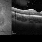
 Serpiginous Choroidal Atrophy
Mar 29 2019 by Jessica Norkus
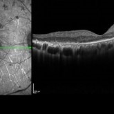
Heidelberg single vertical scan image of 20-year-old female presenting with serpiginous choroidal atrophy. Patient was unaware of vision loss OD, until accidentally covering OS and noticing the change. Acuity of 20/200 OD and 20/15 OS at time of imaging.
Photographer: Jessica Norkus
Imaging device: Heidelberg Spectralis
Condition/keywords: Heidelburg Spectralis, macula lesion, macula serpiginous choroidopathy, optical coherence tomography (OCT)
-
 Pigmentary Retinal Dystrophy
Mar 29 2019 by Jessica Norkus
Heidelberg Spectralis image of 41-year-old male patient with pigmentary retinal dystrophy. Atypical findings due to unilateral presentation. Patient has been experiencing symptoms for 15 years, notes significant nyctalopia.
Photographer: Jessica Norkus
Imaging device: Heidelberg Spectralis
Condition/keywords: bone spicule, Heidelburg Spectralis, optical coherence tomography (OCT), pigment changes, unilateral blindness
 Loading…