 Initializing download.
-
 By samarth mishra
Sankara Nethralaya
Co-author(s): Dr Kumar Saurabh - Uploaded on Sep 25, 2018.
- Last modified by Caroline Bozell on Sep 25, 2018.
- Rating
- Appears in
- Miscellaneous
- Condition/keywords
- multicolor, optical coherence tomography (OCT), Heidelburg Spectralis, diabetic retinopathy
- Photographer
- Aditya Birla Sankara Nethralaya, Kolkata, West Bengal , India
- Imaging device
- Scanning laser ophthalmoscope
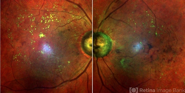
- Description
- A 60-year-old male presented with a history of blurring of vision since many months. He had a history of diabetes since last 8 years. On routine examination proliferative diabetic retinopathy with diabetic macular edema was noted. Fundus fluorescein angiography showed neovascularization elsewhere. Hard exudates can be seen as greenish yellow dots all over the posterior pole in multicolor imaging. Retinal hemorrhage can be seen as dark red.

")


")
")
")


