 Initializing download.
-
 By samarth mishra
Sankara Nethralaya
Co-author(s): Dr Rupak Roy, Dr Kumar Saurabh - Uploaded on Sep 25, 2018.
- Last modified by Caroline Bozell on Sep 25, 2018.
- Rating
- Appears in
- Miscellaneous
- Condition/keywords
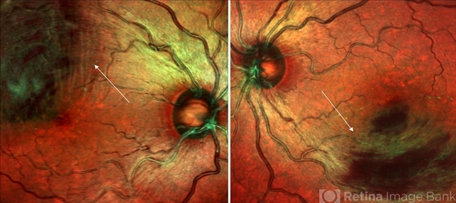
- multicolor, optical coherence tomography (OCT), Heidelburg Spectralis, branch retinal vein occlusion (BRVO)
- Photographer
- Aditya Birla Sankara Nethralaya, Kolkata, West Bengal , India
- Imaging device
- Optical coherence tomography system
- Description
- A 40-year-old female presented with complains of blurring of vision since past 1 week. Patient had a history of hypertension. On routine examination bilateral branch retinal vein occlusion was noted. Visual acuity at presentation was 6/9 and 6/15 in the right and left eye respectively. Multicolor composite imaging shows the hemorrhage as red and the retinal thickening as greenish hue. She was managed with anti vascular endothelial growth factor in both eyes.


")


 Caused due Branch Retinal Vein Occlusion (BRVO)")