Search results (189 results)
-
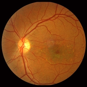
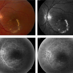
 Ocular Toxocariasis slide 1
Oct 22 2012 by Ronald C. Gentile, MD
8-year-old boy with a history of puppy exposure failed his school screening in the right eye. Fundus examination revealed a old scarred granuloma involving the macula. Serum testing for anti-Toxocara antibodies were positive.
Photographer: The New York Eye & Ear Infirmary Department of Medical Imaging
Condition/keywords: scarred granuloma, toxocariasis
-
000---thumb.jpg/image-square;max$300,300.ImageHandler) Fundus Panorama Finding of Tractional Retinal Detachment Due to Proliferative Diabetic Retinopathy
Fundus Panorama Finding of Tractional Retinal Detachment Due to Proliferative Diabetic Retinopathy
Dec 25 2013 by Dong Yoon Kim, MD
47-year-old woman visited our clinic for decreased visual acuity on her right eye. Her visual acuity of right eye was hand motion. Fundus examination showed traction retinal detachment.
Condition/keywords: fundus photograph, tractional retinal detachment
-
---thumb.jpg/image-square;max$300,300.ImageHandler) Birdshot Choroidopathy
Birdshot Choroidopathy
Oct 9 2013 by Maurice F. Rabb
Forty two year old white female first noted flashing lights in her left eye at the age of 30. Although she had many previous eye examinations for low grade myopia, she had never had a dilated fundus examination. The evaluation twelve years ago disclosed 20/20 acuity in each eye with a myopic correction, an afferent pupillary defect on the left, no evidence of anterior segment inflammation in either eye, a full field on the right and markedly constricted field on the left, fundus pigmentary abnormalities extending beyond the equator in each eye, and narrow vessels with pigment migration into the retina in the left eye only.
Condition/keywords: birdshot choroidopathy
-
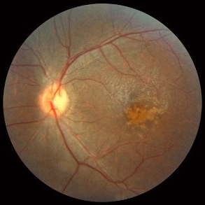
 Stargardt macular dystrophy slide 2
Oct 22 2012 by Ronald C. Gentile, MD
Fundus examination of the left eye had similar findings with centrally atrophic macula area with surrounding flecks.
Photographer: The New York Eye & Ear Infirmary Department of Medical Imaging
Condition/keywords: Stargardt disease
-
---thumb.jpg/image-square;max$300,300.ImageHandler) OCT-CMV-Macula-SRF
OCT-CMV-Macula-SRF
Feb 24 2014 by Susanna S. Park, MD, PhD
Macular OCT of a 59-year-old woman on systemic chemotherapy for acute lymphocytic leukemia with new vision loss. Macular infiltration and submacular fluid are noted. Fundus examination showed a hemorrhagic retinitis in the macula from cytomegalovirus infection.
Photographer: Karishma Chandra, University of California Davis Eye Center
Imaging device: Cirrus OCT
Condition/keywords: CMV retinitis, optical coherence tomography (OCT)
-
 Toxoplasmosis Slide 1
Oct 22 2012 by Ronald C. Gentile, MD
35-year-old women presented with decreasing vision in the left eye with progressive central scotoma. Fundus examination revealed one focal area of chorioretinitis adjacent to one of multiple old pigmented retinal scars. The focal area of chorioretinitis involved the deep retinal layers and was associated with sub-retinal fluid and little overlying vitritis.
Photographer: The New York Eye & Ear Infirmary Department of Medical Imaging
Condition/keywords: punctate outer retinal toxoplasmosis, toxoplasmosis
-
 cRORA
cRORA
Aug 5 2020 by Dhaivat Shah
A 54-year-old healthy male presented to us with a decreased vision in right eye since past 8 years. The patient gave a history of bleed in right eye before 8 years for which some intravitreal injection was given; post which there no major visual improvement. No details or documentation was available regarding the same. His BCVA in the right eye was 5/60. Fundus examination revealed a sharply demarcated hypopigmented patch over the macula with mild posterior excavation suggestive of macular scar. OCT image shows foveal thinning with loss of Retinal pigment epithelium and outer retinal layers (RORA). There are 2 types of RORAs, complete and incomplete. Complete RORA and incomplete RORA are entities defined by various imaging modalities describing atrophy of the retinal pigment epithelial and the outer retinal layers. OCT imaging defines incomplete RORA (iRORA) as a region of signal hyper transmission into the choroid and a corresponding zone of attenuation ordisruption of the RPE (<250um) and evidence of overlying photoreceptor degeneration (<250um). There should not be any RPE tear associated with it. OCT imaging describes complete RORA (cRORA) based on 4 inclusion criteria. These include, area of hypertransmission of more than 250um, zone of attenuation or disruption of the RPE of more than 250um in diameter, evidence of overlying photoreceptor degeneration and absence of scrolled RPE or other signs of an RPE tear. Other modalities used to define these include fundus autoflourescence(FAF), near infrared reflectance(NIR) and color fundus photograph(CFP). On CFP, it shows a sharply demarcated hypopigmented of >250um size with better visibility of choroidal vessels. FAF shows a hypo autoflourescent patch with sharply demarcated borders of size >250um, the colour of which is similar to that of the optic nerve head or retinal blood vessels excluding any pigmentation or artefact. On NIR, it shows a hyperreflective area with sharply demarcated borders of >250um size excluding any artefact. RORA can be seen in conditions like geographical atrophy in ARMD, central areolar choroidal dystrophy, atrophy secondary to anti-VEGF treatment. References: 1. Sadda SR, Guymer R, Holz FG, et al. Consensus Definition for Atrophy Associated with Age-Related Macular Degeneration on OCT: Classification of Atrophy Report 3 [published correction appears in Ophthalmology. 2019 Jan;126(1):177]. Ophthalmology. 2018;125(4):537-548. 2. Guymer RH, Rosenfeld PJ, Curcio CA, et al. Incomplete Retinal Pigment Epithelial and Outer Retinal Atrophy in Age-Related Macular Degeneration: Classification of Atrophy Meeting Report 4. Ophthalmology. 2020;127(3):394-409. 3. Eng VA, Rayess N, Nguyen HV, Leng T. Complete RPE and outer retinal atrophy in patients receiving anti-VEGF treatment for neovascular age-related macular degeneration. PLoS One. 2020;15(5):e0232353.
Photographer: Miss Anjum Zafar Khan
Imaging device: Choithram Netralaya
Condition/keywords: macular scar, outer retina, retinal pigment epithelium
-
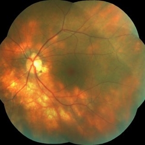
 Cone Rod Dystrophy slide 2
Oct 22 2012 by Ronald C. Gentile, MD
Fundus examination revealed spotty pigmentary changes in the macular area with some peripheral depigmentation of the retinal pigment epithelium.
Photographer: The New York Eye & Ear Infirmary Department of Medical Imaging
Condition/keywords: cone dystrophy, retinal pigment epithelium
-
---thumb.jpg/image-square;max$300,300.ImageHandler) Birdshot Choroidopathy
Birdshot Choroidopathy
Oct 9 2013 by Maurice F. Rabb
Forty two year old white female first noted flashing lights in her left eye at the age of 30. Although she had many previous eye examinations for low grade myopia, she had never had a dilated fundus examination. The evaluation twelve years ago disclosed 20/20 acuity in each eye with a myopic correction, an afferent pupillary defect on the left, no evidence of anterior segment inflammation in either eye, a full field on the right and markedly constricted field on the left, fundus pigmentary abnormalities extending beyond the equator in each eye, and narrow vessels with pigment migration into the retina in the left eye only.
Condition/keywords: birdshot choroidopathy
-
 Anterior ischemic optic neuropathy slide 1
Oct 22 2012 by Ronald C. Gentile, MD
70-year-old women with acute loss of vision in the left eye. Review of symptoms was significant for temporal arteritis and ESR was very high. Fundus examination of the left eye had a swollen white optic nerve head with a few peri-papillary cotton wool spots.
Photographer: The New York Eye & Ear Infirmary Department of Medical Imaging
Condition/keywords: anterior ischemic optic neuropathy, choroidal ischemia, temporal arteritis
-
 Valsalva Retinopathy
May 30 2014 by Mitzy E Torres Soriano, MD
A 45-year-old woman presented sudden loss of vision (hand motion) in the right eye. Fundus examination revealed multiple deep retinal hemorrhages and a large pre macular subhyaloid hemorrhage. Spontaneous resorption occurred at one month and visual acuity improved to 20/25.
Photographer: Mitzy E Torres Soriano. Hospital Central de Maracay. Venezuela
Condition/keywords: macular hemorrhage, subhyaloid hemorrhage, valsalva retinopathy
-
 Ocular Hypotony Due to Leaking Bleb
Apr 1 2019 by Anfisa Ayalon, MD
81-year-old male who had trabeculectomy in his right eye 4 years ago, presented to the emergency room with complains of decreased vision in that eye for two months. Slit-lamp examination showed cystic bleb with leakage, intraocular pressure was 0 MMHg. Fundus examination showed hypotony maculopathy, peripheral choroidal detachments, multiple chorioretinal folds with subretinal fluid.
Photographer: Anfisa Ayalon, MD., Meir Medical Center, Kfar Saba, Israel.
Imaging device: California, Optos 200 DTX
Condition/keywords: choroidal detachment, hypotonous retinopathy, hypotony maculopathy
-
 Chorioretinitis Sclopetaria
Jan 22 2016 by Jorge Morales-Martínez, MD
Fundus photograph of a 27-year-old male that sustained a traumatic injury in his left eye with a paintball projectile. Fundus examination showed a large subretinal hemorrhage, areas of commotio retinae and maculopathy.
Photographer: Jorge Morales-Martínez MD
-
 Hemangioma
Oct 16 2012 by Anat Loewenstein, MD
Fundus examination of a 68 year old lady with decreased vision in her left eye for several months. VA 20/30 in her RE and counting fingers in the left eye. In the left eye there was a large red mass protruding and covering almost the entire optic nerve. Diagnosed as retinal hemangioma. The patient underwent low fluence PDT.
Photographer: Galit Yair-Pur
Condition/keywords: hemangioma
-
 Dislocated Lens
Jun 29 2013 by Jason S. Calhoun
84-year-old female comes in with blurred vision in the left eye. VA was 20/30, right eye and count fingers in the left eye. Fundus examination reveals dislocation of the IOL into the vitreous inferiorily at 6-o'clock. Suggest surgery to fix the problem.
Photographer: Jason S. Calhoun, Mayo Clinic Jacksonville, Florida
Imaging device: TOPCON TRC 50-EX
Condition/keywords: dislocated posterior chamber intraocular lens (PCIOL)
-
 Birdshot Case #1 OS Color
May 1 2013 by Armando L. Oliver, MD
64-year-old Puerto Rican woman consulted due to the presence of 1+ vitreous cells. The fundus examination revealed orange to yellow lesions dispersing from the disk. Work-up revealed she was HLA-A29 positive and the suspected diagnosis of Birdshot Chorioretinopathy was made. Chest X-Ray, FTA-Abs and RPR were negative.
Photographer: Moises Castro, Instituto de Ojos y Piel, Carolina, PR
Imaging device: Zeiss, Visucam NM/FA
Condition/keywords: birdshot, birdshot chorioretinopathy, birdshot retinochoroidopathy
-
---thumb.jpg/image-square;max$300,300.ImageHandler) Birdshot Case #1 OD Color
Birdshot Case #1 OD Color
May 1 2013 by Armando L. Oliver, MD
64-year-old Puerto Rican woman consulted due to the presence of 1+ vitreous cells. The fundus examination revealed orange to yellow lesions dispersing from the disk. Work-up revealed she was HLA-A29 positive and the suspected diagnosis of Birdshot Chorioretinopathy was made. Chest X-Ray, FTA-Abs and RPR were negative.
Photographer: Moises Castro, Instituto de Ojos y Piel, Carolina, PR
Imaging device: Zeiss, Visucam NM/FA
Condition/keywords: birdshot, birdshot chorioretinopathy, birdshot retinochoroidopathy
-
 Astrocytic Hamartoma
Apr 30 2015 by Mariam A Al-Feky, MD
A 15-year-old boy with history of seizures controlled on treatment. C/O: OD painless DV 10/7 ago (accidental discovery) O/E: BCVA OD: 6/60 ,, OS 6/6. AS: NAD OU. Pupil: RRR no RAPD OU. Fundus examination OD showed a retinitis like lesion with an overlying corkscrew vessel well evident on FFA with late leakage and CSR and OCT through the retinitis like lesion shows diffuse hypereflective thickeninig in the superficial NFL. Thorough history taking revealed that patient has seizures and MRI lesions suggestive of tuberous sclerosis. So this is exudative hamartoma secondary to tuberous sclerosis with marked resolution after single IVI of Lucentis. Retinitis like lesion with corkscrew vessels in FFA is typical together with the homogenous hypereflective thickening in the NFL.
Photographer: Mariam AL-Feky
Imaging device: Optical coherence tomography
Condition/keywords: astrocytic hamartoma
-
---thumb.jpg/image-square;max$300,300.ImageHandler) Multiple Vitelliforn Retinal 6
Multiple Vitelliforn Retinal 6
Mar 15 2013 by Maurice F. Rabb
44-year-old female patient on a return visit after being diagnosed with Sjogren's syndrome with swelling of the parotid glands. Fundus examination showed resolution of much of the subretinal yellow material previously found. Both foveal areas were flat with pigmentary mottling. There were still remnants of yellowish material outside of the fovea.
Condition/keywords: fovea, subretinal, vitelliform lesion
-
---thumb.jpg/image-square;max$300,300.ImageHandler) Birdshot Choroidopathy
Birdshot Choroidopathy
Oct 9 2013 by Maurice F. Rabb
Forty two year old white female first noted flashing lights in her left eye at the age of 30. Although she had many previous eye examinations for low grade myopia, she had never had a dilated fundus examination. The evaluation twelve years ago disclosed 20/20 acuity in each eye with a myopic correction, an afferent pupillary defect on the left, no evidence of anterior segment inflammation in either eye, a full field on the right and markedly constricted field on the left, fundus pigmentary abnormalities extending beyond the equator in each eye, and narrow vessels with pigment migration into the retina in the left eye only.
Condition/keywords: birdshot choroidopathy
-
---thumb.jpg/image-square;max$300,300.ImageHandler) Multiple Vitelliforn Retinal 5
Multiple Vitelliforn Retinal 5
Mar 15 2013 by Maurice F. Rabb
44-year-old female patient on a return visit after being diagnosed with Sjogren's syndrome with swelling of the parotid glands. Fundus examination showed resolution of much of the subretinal yellow material previously found. Both foveal areas were flat with pigmentary mottling. There were still remnants of yellowish material outside of the fovea.
Condition/keywords: fovea, subretinal, vitelliform lesion
-
---thumb.jpg/image-square;max$300,300.ImageHandler) Multiple Vitelliforn Retinal 7
Multiple Vitelliforn Retinal 7
Mar 15 2013 by Maurice F. Rabb
44-year-old female patient on a return visit after being diagnosed with Sjogren's syndrome with swelling of the parotid glands. Fundus examination showed resolution of much of the subretinal yellow material previously found. Both foveal areas were flat with pigmentary mottling. There were still remnants of yellowish material outside of the fovea.
Condition/keywords: fovea, subretinal, vitelliform lesion
-
 Vascular Anormalities
Jan 6 2016 by Andrea Arriola-Lopez, MD MSc
77-year-old man. Decrease of visual acuity OS. VA 20/30 IOP 14mmHg. Fundus examination findings: Hard exudates, microaneurysms near to fovea. OCT shows IRF. Late leakage on FA.
Photographer: Andrea Elizabeth Arriola-Lopez, MSc MD
Condition/keywords: abnormal retinal vessel, aneurysm, hard exudates, vascular anomaly
-
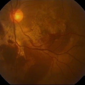
 Chronical Submacular Hemorrhage in the Setting of Neovascular AMD
Mar 23 2015 by Rita Couceiro, MD, MS
An 80-year-old male, with a history of hypertension and high cholesterol, complained of acute and painless vision loss in his left eye (OS) in the previous 5 months. On observation best corrected visual acuity in OS was hand motion. A dense vitreous opacity in OS precluded fundus examination. Ocular ultrasound revealed vitreous hemorrhage and thickening of the macular area. The patient was submitted to pars plana vitrectomy, which disclosed a large submacular hemorrhage with chronical features and disciform scarring in the setting of neovascular AMD.
Imaging device: Intraoperative fundus photograph
Condition/keywords: neovascular age-related macular degeneration (AMD), submacular hemorrhage, wet age-related macular degeneration (wet AMD)
-
---thumb.jpg/image-square;max$300,300.ImageHandler) Birdshot Case #1 OD IVFA
Birdshot Case #1 OD IVFA
May 1 2013 by Armando L. Oliver, MD
64-year-old Puerto Rican woman consulted due to the presence of 1+ vitreous cells. The fundus examination revealed orange to yellow lesions dispersing from the disk. Work-up revealed she was HLA-A29 positive and the suspected diagnosis of Birdshot Chorioretinopathy was made. Chest X-Ray, FTA-Abs and RPR were negative.
Photographer: Moises Castro, Instituto de Ojos y Piel, Carolina, PR
Imaging device: Zeiss, Visucam NM/FA
Condition/keywords: birdshot, birdshot chorioretinopathy, birdshot retinochoroidopathy
 Loading…