Search results (800 results)
-
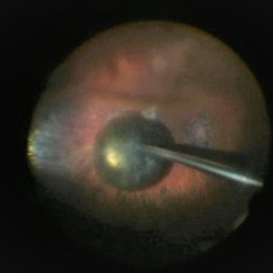
 Foreign Body SS
Feb 14 2015 by Shlomit Schaal, MD, PhD, MHCM
A four millimeter metal foreign body removed surgically using foreign body forceps. The macula is protected by PFCL heavy fluid. There is a traumatic retinal detachment inferiorly (top of photo, surgeon's view).
Photographer: Shlomit Schaal, MD, PhD
Condition/keywords: intraocular foreign body
-
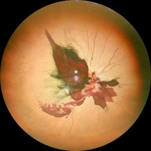
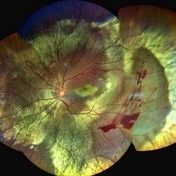
 Post Traumatic Optic Nerve Head Avulsion
Nov 18 2017 by Vishal Agrawal, MD, FRCS,FACS,FASRS
Right eye fundus picture of a 24-year-old male patient who suffered blunt trauma 7 days back with a wooden stick . He presented with NLP vision with a non reacting dilated pupil. Fundus montage picture shows ONH avulsion,CRAO,peripapillary resolving hemorrhages and cicatricial tissue at the edge.
Photographer: Vishal Agrawal, MD, SMS Medical College, Jaipur, India
Imaging device: Zeiss 524
Condition/keywords: avulsion, central retinal artery occlusion (CRAO)
-
 Giant Retinal Tear
Feb 20 2024 by Soobien Lee
Optos color fundus photograph of a 40-year-old caucasian male who is a UFC fighter with a total retinal detachment in his right eye secondary to a giant retinal tear from 10 o'clock to 2 o'clock.
Photographer: Trinity Wolf, Elman Retina Group
Imaging device: Optos Ultra-Widefield Imaging
Condition/keywords: giant retinal tear, optos, Retinal Detachment, Retinal tear with detachment, trauma
-
 Optic Nerve Avulsion with Vitreous Hemorrhage and Pale Retina
Jan 25 2021 by Sham Talati, DOMS
A 30-year-old male presented with history of trauma to RE with NO Perception of light in the affected eye.
Photographer: Dr. Sham Talati,Retina Foundation,Ahmedabad
Imaging device: Nidek Mirante
Condition/keywords: optic nerve, pale retina
-
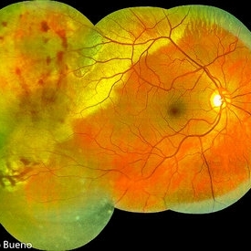 Commotio Post-Blunt Trauma
Apr 3 2018 by Paulo Bueno
Fundus photograph of an 35-year-old man with commotio retinae after indoor soccer ball blunt trauma.
Photographer: Paulo Bueno, Taubaté, Brazil.
Imaging device: Zeiss Visucam
Condition/keywords: blunt trauma, commotio retinae
-
 Central Retinal Vein Occlusion
Jan 21 2022 by Olivia Rainey
Ultra-widefield fluorescein angiogram of a 23-year-old female with a Central Retinal Vein Occlusion affecting her left eye. The patient presented on 12/22/2021 cc20/40-2 vision in the left eye. The patient reported recent trauma of being hit with a fist on both sides of face followed by vision loss. The patient has history of Hashimoto's thyroid disease. The following labs have been ordered, PT, PTT, CBC, antithrombin III activity, protein C, protein S, Factor V Leiden mutation, Prothrombin (G20210A), lipid panel, HbA1c, quantiferon gold, RPR, and CXR.
Photographer: Olivia Rainey, OCT-C, COA
Imaging device: Optos California
Condition/keywords: central retinal vein occlusion (CRVO), disc leakage, fluorescein angiogram (FA), fluorescein leakage, left eye, non-ischemic central retinal vein occlusion (CRVO), Optos, trauma, ultra-wide field imaging
-
 Choroidal Fracture
Oct 27 2024 by César Adrián Gómez Valdivia, MD
Fundus photograph of a traumatic choroidal fracture & extra-macular sub-retinal hemorrhage.
Photographer: @eyemissu2
Imaging device: TOPCON TRC-50DX
Condition/keywords: Choroidal Fracture
-
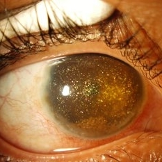
 Dropped Crystalline Lens
Mar 8 2019 by Abdulaziz A. Alshamrani, MD
A 15-year-old female with congenital glaucoma complaining of acute diminution of vision after a blunt trauma.
Condition/keywords: crystalline lens, dropped nucleus, ora serrata
-
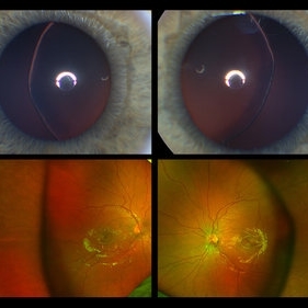 Ectopia Lentis
Jan 21 2021 by Jamin S. Brown, MD
This image serial demonstrates a patient with simple ectopia lentis. Anterior segment photographs in the upper panel demonstrate nasally subluxated crystalline lenses. Widefield fundus photography shows a "pseudo-buckle" which is the result of an optical effect due to the lens subluxation (artifactual image enlargement). Also note the juvenile macular reflex in this young patient. Ectopia lentis can present isolated ("simple") or in combination with various systemic defects (Marfan's syndrome, Weil-Marchesani syndrome or Ehlers-Danlos syndrome to name a few). Isolated ectopia lentis can be hereditary and causative genes have been identified as ADAMTSL4 located on chromosome 4 and FBN1 gene located on chromosome 15. Defects in the genes cause weakness in the zonular fibers which can lead to lens dislocation. Lastly, various ocular disorders such as Aniridia, Axenfeld-Rieger, Pseudoexfoliation or Trauma may also result in lens dislocation or subluxation.
Photographer: Stefanie Palmer CRA, Retina Vitreous Surgeons of CNY
Condition/keywords: dislocated lens, ectopia lentis
-
 Fraternal Twins
May 22 2023 by Gustavo M. Hüning, MD, MBA, FASRS
Intrasurgical photograph using a non-contact system and 3D visualization system of a 65-year-old woman who suffered an ocular trauma.
Photographer: Gustavo M. Hüning, Hüning Clínica do Olhar, Santa Maria - Brazil
Imaging device: Alcon Luxor combined with Alcon nGenuity
Condition/keywords: dislocated intraocular lens (IOL), implant, pars plana vitrectomy (PPV)
-
 Massive Commotio Retinae
Oct 20 2020 by Veronika Yehezkeli
Fundus photograph of a 24-year-old male, made after blunt trauma with a plastic bottle. Note massive commotio retinae and preretinal hemorrhages in the contralateral to trauma area.
Photographer: Veronika Yehezkeli, Meir medical center, Israel
Condition/keywords: blunt trauma, commotio retinae, preretinal hemorrhage, trauma
-
 Optic disc avulsion
Mar 5 2023 by Kalyan Singh
Case of post traumatic optic disc avulsion of young male adult.
Photographer: Kalyan Singh, GSVM medical college, Kanpur
Imaging device: Smartphone (1 plus 10R)
Condition/keywords: optic disc, trauma
-
 Optic Nerve Head Avulsion
Sep 24 2024 by Gustavo Uriel Fonseca Aguirre
A 14-year-old male with a history of blunt ocular trauma in the right eye presented partial avulsion of the optic nerve head and submacular hemorrhage that was managed with neumatic displacement.
Photographer: Gustavo U. Fonseca Aguirre, Fundación Hospital Nuestra Señora de la Luz, Ciudad de México
Condition/keywords: optic nerve head avulsion
-
 PPV retained cataract
Apr 19 2023 by Denica Rodriguez
A 46-year-old male with hypermature dense cataract. Patient got a piece of metal in his eye when he was 5 years old and was not able to see since. Patient was having cataract surgery and phacodonesis was present. The lens dropped to the back of the eye. Patient had to have another surgery to do vitrectomy. The lens removal was done with a fragmatome handpiece.
Photographer: Denica Rodriguez COA, ST
Imaging device: Zeiss Microscope with resight
Condition/keywords: cataract, dropped nucleus, fragmatome, lens capsule, ocular trauma, pars plana vitrectomy (PPV), retained lens fragments, Retina, retina surgery, traumatic cataract
-
 Sympathetic Ophthalmia
Jul 12 2021 by Stefanie Palmer
Fundus photo of a 57-year-old man with history of trauma to the fellow eye.
Photographer: Stefanie Palmer, CRA
Condition/keywords: sympathetic ophthalmia, sympathetic uveitis
-
 Synchysis Scintillans
Sep 17 2015 by Jessica G Lee, MD
24-year-old male with history of chronic retinal detachment.
Photographer: Bob Masini
Condition/keywords: cholesterol crystals, refractile bodies, synchysis scintillans, trauma, vitreous hemorrhage
-
 Traumatic Choroidal Rupture
Jun 11 2017 by Bastián Schmidt Arias
Fundus photograph of an 28-year-old men with a traumatic choroidal rupture. Visual acuity is CF 30 cm.
Photographer: Bastian Schmidt
Imaging device: TRC-50DX - Topcon
Condition/keywords: choroidal rupture, fundus photograph
-
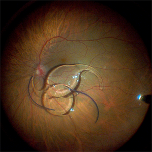
 Traumatic perforation and PVR
May 19 2022 by ALLAN GOMES DA SILVA
PVR, retinal detachment and macular dragging after iatrogenic traumatic perforation during peribulbar block. We can observe the inferior perforation, the brutal formation of PVR and the fovea being pulled.
Photographer: Edimilson Ferreira da Silva
Imaging device: Topcon TRC-50 DX, Imaginet 4.0, angle - 50 degrees
Condition/keywords: posterior perforation, proliferative vitreoretinopathy (PVR)
-
 Traumatic Retinal Tear
Dec 5 2021 by Aditya S Kelkar, MS, FRCS, FASRS,FRCOphth
Color fundus photograph of a 34-year old man's left eye, 2 hours after a tennis ball injury, showing commotio retinae with Berlin's edema and cherry red spot in the fovea along with linear retinal tears in the temporal equatorial zone.
Photographer: Dr Sukanya Mondal. National Institute of Ophthalmology, Pune. India.
Imaging device: Zeiss Clarus 500
Condition/keywords: Berlin's edema, cherry red spot, commotio retinae, retinal tear
-
 4 Point Scleral Fixation Akreos AO60 With Gore Tex Suture
May 21 2021 by Jesus Lozano, MD
Anterior segment photo of a 54-year-old man after 4 point scleral fixation Akreos AO60 with Gore Tex suture plus PPV who had a severe traumatic iris defect and was aphakic after ocular trauma.
Photographer: Luigi Zinn, Hadassah Medical Center, Jerusalem.
Condition/keywords: aphakia, cornea rupture, lens, penetrating trauma
-
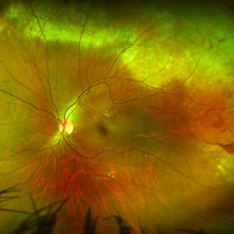
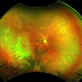
 A Large Break at the Posterior Pole With RD With PVR (S/p Old Blunt Trauma)
Jan 16 2025 by Anand Temkar
Right eye central fundus color photo of a 10 year old kid who noticed diminution of vision in right eye since a month. We can see the large break at the posterior pole with rolled up margins associated with retinal detachment and PVR changes.
Photographer: Dr.Anand Temkar- Retina Foundation, Ahmedabad
Imaging device: Mirante
Condition/keywords: Posterior pole break, proliferative vitreoretinopathy (PVR), Retinal Detachment
-
 Angioid streaks - PXE case 2
Jan 11 2013 by Alex P. Hunyor, MD
Pseudoxanthoma elasticum with angioid streaks, left eye - note haemorrhages along angioid streaks following minor blunt ocular trauma.
Condition/keywords: angioid streaks, pseudoxanthoma elasticum (PXE)
-
 Auto-Enucleation with Tire Iron
Oct 19 2012 by Larry Halperin, MD
Auto-enucleation with tire iron
Condition/keywords: enucleation, self-inflicted, trauma
-
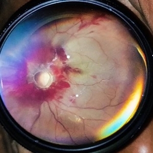
 Choroidal Rupture
Jun 29 2013 by Jason S. Calhoun
Adult male with trauma to the right eye and orbital floor fracture. Hemorrhage with choroidal rupture.
Photographer: Jason S. Calhoun, Mayo Clinic Jacksonville, Florida
Imaging device: TOPCON TRC 50-EX
Condition/keywords: choroidal rupture
-
 Choroidal Rupture
Sep 30 2023 by Jacob D. Grodsky, MD
24 year old female who presented after being hit in the head with a metal softball bat after an altercation. The patient reported blurred vision as well as a zig-zag line described as a “lightning strike” across her vision. Examination was significant for a choroidal rupture OD as well as commotio retinae OU.
Condition/keywords: choroidal rupture, commotio retinae, trauma
 Loading…