Search results (800 results)
-
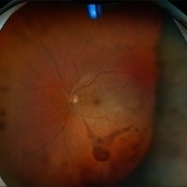
 Traumatic Hemorrhage
Apr 17 2025 by Virginia Gebhart
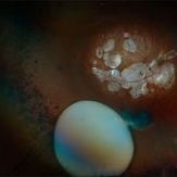
60 year old male with vitreous and sub hyaloid hemorrhage from being hit in the eye. No holes, tears, or detachment. Will observe closely, if no improvement will consider surgical repair. Treated melanoma s/p brachytherapy in 2008.
Photographer: Virginia Gebhart, Retina Consultants of Carolina
Imaging device: Optos California
Condition/keywords: blunt trauma, sub hyaloid hemorrhage, vitreous hemorrhage
-
 Traumatic Posterior Capsular Rupture
Apr 9 2025 by Gustavo Uriel Fonseca Aguirre
Immersion B-mode ultrasound in a patient with blunt ocular trauma demonstrates an isolated posterior lens capsule rupture accompanied by phacodonesis.
Photographer: Gustavo U. Fonseca Aguirre, Hospital Conde de Valenciana, Ciudad de México
Condition/keywords: blunt trauma, Posterior Capsular Rupture
-
 Choroidal Rupture
Apr 7 2025 by Ramses Rosales-Diaz
Autofluorescence image of a 39-year-old female patient who sustained blunt ocular trauma resulting in three choroidal ruptures.
Photographer: Ramses Rosales-Diaz, Asociación Para Evitar la Ceguera en México I.A.P., Mexico City
Imaging device: Heidelberg Spectralis
Condition/keywords: blunt trauma, Choroidal Rupture
-
 Cyclodialysis
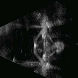
Apr 3 2025 by Gustavo Uriel Fonseca Aguirre
Ultrasound biomicroscopy (UBM) of a blunt-traumatized eye revealing cyclodialysis, zonular disruption with lenticular ectopia, and anterior chamber vitreous prolapse.
Photographer: Gustavo U. Fonseca Aguirre, Hospital Conde de Valenciana, Ciudad de México
Condition/keywords: cyclodialysis
-
 Intraocular Foreign Body
Apr 3 2025 by Gustavo Uriel Fonseca Aguirre
B-mode ultrasonography of an eye with a 1-year history of suspected blunt trauma revealed an incidental intraocular foreign body within the vitreous cavity.
Photographer: Gustavo U. Fonseca Aguirre, Hospital Conde de Valenciana, Ciudad de México
Condition/keywords: intraocular foreign body
-
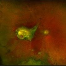
 Bleb Migration With FAX
Mar 25 2025 by Robert Andrew Sisk, MD, FACS, FASRS
Color stills from surgical video after subretinal delivery of gene augmentation therapy with voretigene neparvovec-rzyl A) before and B) after fluid-air exchange (FAX). The blebs were between 0.5- and 1-disc diameters from the fovea. After FAX, they gradually extended beneath the fovea and eventually merged. This spared the fovea the trauma from the injection pressure of subretinal injection while allowing treatment to the area.
Imaging device: Leica Proveo 8
Condition/keywords: Fluid-Air Exchange, Gene Therapy, genetic disorder, genetics, Subretinal Injection
-
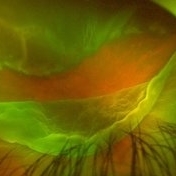
 Firework Injury
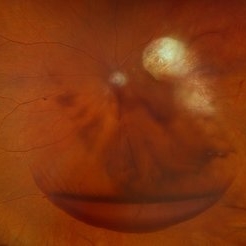
Feb 13 2025 by Virginia Gebhart
44 year old male presented New Year's Day for trauma after fireworks injury. Choroidal rupture temporal macula, inferior vitreous hemorrhage, and extensive RPE changes in the macula. Significant improvement since initial presentation. Limited central vision, guarded prognosis due to extensive blunt trauma.
Photographer: Virginia Gebhart, Retina Consultants of Carolina
Imaging device: Optos California
Condition/keywords: blunt trauma, choroidal rupture, commotio retinae, firework injury, secondary glaucoma, subretinal hemorrhage, VH, vitreous hemorrhage
-
 Dislocated Lens
Jan 30 2025 by Kimberly Wakester
Fundus photograph of a 37-year-old man with an anteriorly dislocated lens in the left eye. The natural lens has displaced anteriorly in the AC secondary to trauma to the eye. There is also a Macular hole present with vitreous hemorrhage. Patient was recommended to proceed with lensectomy, iris repair and MH repair in the left eye.
Photographer: Kimberly Wakester, COA
Imaging device: Topcon TRC-50DX
Condition/keywords: dislocated lens, iridodialysis
-
 Macular Hole
Jan 30 2025 by Kimberly Wakester
Fundus photograph of a 37-year-old man with an anteriorly dislocated lens in the left eye. The natural lens has displaced anteriorly in the AC secondary to trauma to the eye. There is also a Macular hole present with vitreous hemorrhage. Patient was recommended to proceed with lensectomy, iris repair and MH repair in the left eye.
Photographer: Kimberly Wakester, COA
Imaging device: Optos California
Condition/keywords: dislocated lens, macular hole, vitreous hemorrhage
-
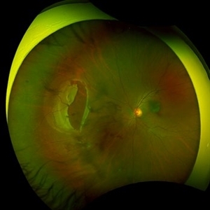
 A Large Break at the Posterior Pole With RD With PVR (S/p Old Blunt Trauma)
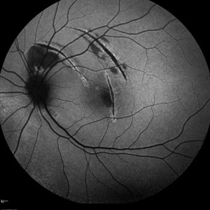
Jan 16 2025 by Anand Temkar
Right eye widefield fundus color photo of a 10 year old kid who noticed diminution of vision in right eye since a month. We can see the large break at the posterior pole with rolled up margins associated with retinal detachment and PVR changes.
Photographer: Dr.Anand Temkar- Retina Foundation, Ahmedabad
Imaging device: Mirante
Condition/keywords: posterior pole break, proliferative vitreoretinopathy (PVR), Retinal Detachment
-
 A Large Break at the Posterior Pole With RD With PVR (S/p Old Blunt Trauma)
Jan 16 2025 by Anand Temkar
Right eye central fundus color photo of a 10 year old kid who noticed diminution of vision in right eye since a month. We can see the large break at the posterior pole with rolled up margins associated with retinal detachment and PVR changes.
Photographer: Dr.Anand Temkar- Retina Foundation, Ahmedabad
Imaging device: Mirante
Condition/keywords: Posterior pole break, proliferative vitreoretinopathy (PVR), Retinal Detachment
-
 Gunshot Injury
Dec 19 2024 by Angela Rico
53 y/o M who suffered gunshot wound to OD. Picture shows macular scar and sub retinal hemorrhage
Photographer: Angela Rico M.D.
Condition/keywords: macular scar, penetrating trauma
-
 Subluxation of the Lens
Dec 12 2024 by Kimberly Wakester
Ultra-wide field fundus photos of an 53-year-old man with a Subluxation of the Lens in the posterior vitreous cavity of the right eye after a trauma that happened many years ago. Patient remains stable with no adverse reaction to the lens at this time. No surgical intervention is recommended at this time. Patient also has myopic degeneration and lattice degeneration that will require patient to have follow up care.
Photographer: Kimberly Wakester, COA
Imaging device: Optos California
Condition/keywords: lattice degeneration, myopic degeneration, peripapillary atrophy, posterior staphyloma, Subluxation of the Lens
-
 Suprachoroidal Hemorrhage
Dec 3 2024 by Dibya Prabha
Colour Fundus photograph of 62 Year old female patient with Suprachoroidal hemorrhage post trauma
Photographer: Dibya Prabha, LV Prasad eye Institute, Hyderabad
Condition/keywords: suprachoroidal hemorrhage
-
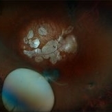
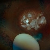
 Traumatic Macular Hole pre and post repair
Nov 25 2024 by Shobhit Chawla, M.S.
31 year-old male reported with h/o of blunt trauma over right eye ,from cricket ball. On examination DVA RE 6/60,LE 6/18,ant segment BE :WNL,FUNDUS RE:Sub retinal hemorrhage at macula with chroidal tear,LE :WNL. Undwer went 25G vitrectomy+sub retinal TPA+C3F8(RE).Post op 1 month DVA RE:6/24 ,ANT SEGMENT:WNL,FUNDUS:resolved sub retinal haem with traumatic macular hole. Under went repeat vit+autologous retinal transplant +SOI RE.POST SOR AFTER4monthsV/A :6/18 RE
Photographer: Ranjit Ray
Imaging device: Clarus 500
Condition/keywords: Macular hole, retinal graft, subretinal hemorrhage
-
 Giant Tear
Oct 28 2024 by Andreas Paulo Di Luciano Rojas, MD
Giant retinal tear secondary to trauma.
Photographer: Andreas Di-Luciano, MD
Imaging device: Optos
Condition/keywords: giant retinal tear, ocular trauma, proliferative vitreoretinopathy (PVR), retinectomy, Trauma
-
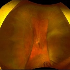
 Choroidal Fracture
Oct 27 2024 by César Adrián Gómez Valdivia, MD
Fundus photograph of a traumatic choroidal fracture & extra-macular sub-retinal hemorrhage.
Photographer: @eyemissu2
Imaging device: TOPCON TRC-50DX
Condition/keywords: Choroidal Fracture
-
 Expulsion of Retina
Oct 23 2024 by Gustavo Uriel Fonseca Aguirre
Male patient with a history of penetrating keratopathy presents due to blunt ocular trauma. A disruption of the continuity at the interface between the donor and recipient corneas is observed, with expulsion of the lens and retina. Vision is limited to light perception.
Photographer: Lizeth Jiménez Santana, Fundación Hospital Nuestra Señora de la Luz, Ciudad de México
Condition/keywords: ocular trauma, penetrating keratoplasty
-
 Large Retinal Tear from a Shuttlecock Injury
Oct 11 2024 by Ahmad B. Tarabishy, MD
27 year old woman presenting with floaters and a shadow in her temporal visual field OS. Approximately one week earlier, she was injured in her left eye by a shuttlecock while playing badminton. Fundus exam reveals mild vitreous hemorrhage and a large retinal tear with a small cuff of surrounding SRF. This image was taken immediately following treatment with barrier laser retinopexy.
Photographer: Angela Rico, M.D.
Imaging device: Optos
Condition/keywords: blunt trauma, ocular trauma, retinal tear
-
 Large Retinal Tear from a Shuttlecock Injury
Oct 11 2024 by Ahmad B. Tarabishy, MD
27 year old woman presenting with floaters and a shadow in her temporal visual field OS. Approximately one week earlier, she was injured in her left eye by a shuttlecock while playing badminton. Fundus exam reveals mild vitreous hemorrhage and a large retinal tear with a small cuff of surrounding SRF.
Photographer: Angela Rico, M.D.
Imaging device: Optos
Condition/keywords: blunt trauma, ocular trauma, retinal tear
-
 Retinitis Pigmentosa With Papilledema
Sep 29 2024 by Tejaswita Verma
Fundus pictures of RE and LE of a 30 year old male with a 20 day history of diminution of vision in LE more than RE. Vision was 6/9P and 6/24P in RE and LE respectively. History of RP in father. Alleged history of trauma to left side of forehead 4 months back with subgaleal haematoma over left side of frontal region on CT done 4 months back, No significant intracranial abnormality. He was started on oral steroids with tapering and F/U after 2 weeks.
Photographer: DR. TEJASWITA VERMA
Imaging device: MIRANTE
Condition/keywords: papilledema, retinitis pigmentosa
-
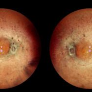
 Optic Nerve Head Avulsion
Sep 24 2024 by Gustavo Uriel Fonseca Aguirre
A 14-year-old male with a history of blunt ocular trauma in the right eye presented partial avulsion of the optic nerve head and submacular hemorrhage that was managed with neumatic displacement.
Photographer: Gustavo U. Fonseca Aguirre, Fundación Hospital Nuestra Señora de la Luz, Ciudad de México
Condition/keywords: optic nerve head avulsion
 Loading…