Search results (799 results)
-
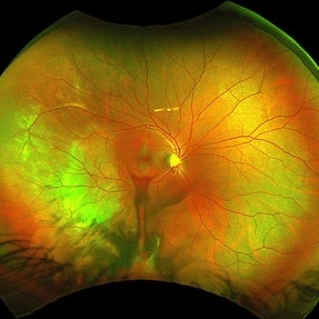
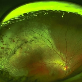
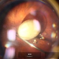
 24 Hours Post Scleral Wound Closure+ Scleral Buckle+25 g Vitrectomy+Silicon Oil
Jan 23 2015 by Carlos Quezada-Ruiz, MD, FASRS
24 hours post op fundus photograph of a 43-year-old man who had perforating injury to the right eye with a small piece of plastic while he was hammering. OD LP, subconjunctival hemorrhage, clear cornea, hyphema, irido and ciclodyalisis as well as a luxated lens with traumatic cataract and a dense vitreous hemorrhage. B-US showed rhegmatogenous retinal detachment with a tear and a big inferior hemorrhagic choroidal detachment. 360 peritomy revealed 2-entry scleral wounds were found in zone II (M V and M VI) and closure was performed. 25 G PPV was performed with the infusion canal placed in the AC through the limbus. Lensectomy and removal of a dense recent vitreous hemorrhage revealed a white detached retina with an exit wound through the temporal inferior segment of the optic nerve with a nasal GRT and sub retinal hemorrhage as well as temporal inferior choroidal, PVD was induced and PFOs helped stabilizing the retina while vitrectomy and sub-retinal hemorrhage was removed through the GRT. Fluid air exchange was made and 360 endolaser over the buckle indentation was done and silicon oil was used as endotamponade. This picture was taken 24 hrs after the surgery.
Photographer: Lilibeth Rodriguez, Instituto de la Visión. Torreon, Mexico.
Condition/keywords: central retinal artery occlusion (CRAO), giant retinal tear, trauma
-
 A Motor Vehicle Accident Causing Valsalva Retinopathy OD, While Racing A Side By Side 4 Wheel Off-Road Vehicle
May 5 2020 by John S. King, MD
A 43-year-old white male who was injured while racing his side by side 4 wheel off-road vehicle (this is a video he showed me on his phone). He presented about three weeks after the injury. He was being seen by his local eye doctor who wanted an evaluation for the retinal heme and scotoma. His main complaint was a central/parcentral scotoma described as a greyish area in vision. Va 20/50 OD, nomotensive, no APD (by technician), anterior segment u/r; see {https://imagebank.asrs.org/file/53828/sxs-crash-during-a-race-causing-valsalva-retinopathy-od} for the fundus exam - of note there are superficial/preretinal heme, with layering of the heme superiorly; in the parafoveal region nasally there is some mottling of the RPE that may indicate an area of prior commotio retinae (also possible to have TON), which may account for his scotoma. Really bad accident, and amazingly, he had no LOC or injuries other than the right retina. Helmet and racing harness seat belt were used.
Condition/keywords: motor vehicle accident, trauma, valsalva retinopathy
-
 Auto-Enucleation with Tire Iron
Oct 19 2012 by Larry Halperin, MD
Auto-enucleation with tire iron
Condition/keywords: enucleation, self-inflicted, trauma
-
 Blunt Trauma
May 18 2016 by Andrea Arriola-Lopez, MD MSc
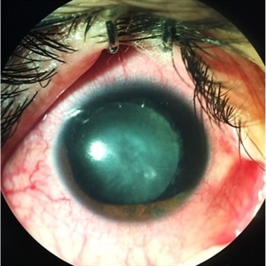
26-year-old man, old ocular blunt trauma. VA HM OD. IOP 14mmHg. Traumatic partial aniridia, cataract and phacodonesys. Ophthalmoscopy showed diffuse hemovitreous, Retina remained attached.
Photographer: Andrea E. Arriola-López MD MSc
Condition/keywords: aniridia, cataract, trauma, traumatic cataract
-
 Blunt Trauma in Angioid Streaks
Jan 5 2018 by Manish Nagpal, MD, FRCS (UK), FASRS
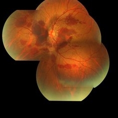
This a Fundus photo of a patient who had come with blunt trauma and on examination had multiple haemorrhages around pre existing angioid streaks.
Photographer: Pooja Barot
Condition/keywords: angioid streaks, trauma
-
 Blunt Trauma in Angioid Streaks
Jan 5 2018 by Manish Nagpal, MD, FRCS (UK), FASRS
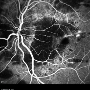
This a fluorescein angiography of a patient who had come with blunt trauma and on examination had multiple haemorrhages around pre existing angioid streaks.
Photographer: Pooja Barot
Condition/keywords: angioid streaks, trauma
-
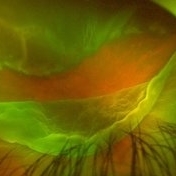
 Central Retinal Vein Occlusion
Jan 21 2022 by Olivia Rainey
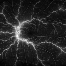
Ultra-widefield fluorescein angiogram of a 23-year-old female with a Central Retinal Vein Occlusion affecting her left eye. The patient presented on 12/22/2021 cc20/40-2 vision in the left eye. The patient reported recent trauma of being hit with a fist on both sides of face followed by vision loss. The patient has history of Hashimoto's thyroid disease. The following labs have been ordered, PT, PTT, CBC, antithrombin III activity, protein C, protein S, Factor V Leiden mutation, Prothrombin (G20210A), lipid panel, HbA1c, quantiferon gold, RPR, and CXR.
Photographer: Olivia Rainey, OCT-C, COA
Imaging device: Optos California
Condition/keywords: central retinal vein occlusion (CRVO), disc leakage, fluorescein angiogram (FA), fluorescein leakage, left eye, non-ischemic central retinal vein occlusion (CRVO), Optos, trauma, ultra-wide field imaging
-
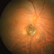
 Choroidal Rupture
Sep 30 2023 by Jacob D. Grodsky, MD
24 year old female who presented after being hit in the head with a metal softball bat after an altercation. The patient reported blurred vision as well as a zig-zag line described as a “lightning strike” across her vision. Examination was significant for a choroidal rupture OD as well as commotio retinae OU.
Condition/keywords: choroidal rupture, commotio retinae, trauma
-
 Choroidal Rupture
Nov 5 2023 by Karen Flores Guevara
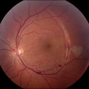
Fundus photograph of a 19-year-old man with a choroidal rupture.
Photographer: Hector Arturo Mendez-Ponce MD, Karen Flores-Guevara MD Asociación para Evitar la Ceguera en México
Condition/keywords: Choroidal, rupture, trauma
-
 Choroidal-rupture
Jan 2 2024 by Tahsin Khundkar, MD
37-year-old male with blunt ocular trauma presented with a choroidal rupture, pre -retinal and sub-retinal heme, and a heart shaped patch of commotio retinae.
Photographer: Jeffrey Zeigler, Concord Eye Center
Imaging device: Topcon
Condition/keywords: Choroidal Rupture, commotio retinae, Trauma
-
 Chronic Intraocular Foreign Body With Siderosis
Jun 28 2014 by John T. Thompson, MD
Large iron containing chronic intraocular foreign body with extensive siderosis of retina. The optic nerve is just to left of foreign body with extensive sheathing of retinal vessels.
Imaging device: Zeiss FF4
Condition/keywords: intraocular foreign body, penetrating trauma, siderosis, trauma
-
 Commotio Retinae
May 19 2014 by John W. Kitchens, MD
Young man with trauma.
Photographer: Ed Slade
Imaging device: Optos 200Tx
Condition/keywords: commotio retinae, trauma
-
 Commotio Retinae
May 19 2014 by John W. Kitchens, MD
Young man with trauma.
Photographer: Ed Slade
Imaging device: Optos 200Tx
Condition/keywords: commotio retinae, trauma
-
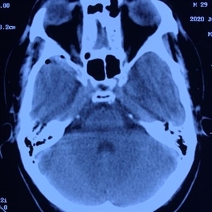 Computed Tomography
Aug 10 2020 by RITESH VERMA
CT scan image section showing fracture of the right greater wing of the sphenoid hampering the blood supply and causing traumatic retinopathy.
Photographer: Dr. Ritesh Verma, Regional institute of Ophthalmology, Rohtak, Haryana, India
Imaging device: CR-2AF CANON
Condition/keywords: CT scan, trauma
-
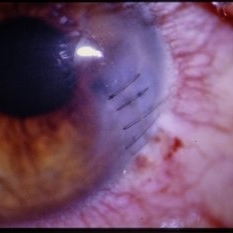 Corneal Wound - Sutured
-
 Crystals in the Eye
Sep 3 2021 by Aditya S Kelkar, MS, FRCS, FASRS,FRCOphth
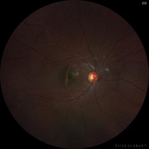
Left eye fundus photo of a 28 year-old , with air-filled vitreous cavity entering through the scleral wound site, after removal of impacted IOFB.
Imaging device: Clarus 500
Condition/keywords: intraocular foreign body, trauma
-
 Dislocated IOL
Jun 4 2024 by Marlee Curnutt
Slit lamp photo of a 64 year old woman presenting with worsening vision and depth perception after trauma induced by a dog, which dislocated her IOL. The patient's IOL haptic was seen in the AC, and almost abutting cornea. Patient's vision upon presentation was DCC CF@1 feet. Patient was counseled and underwent an IOL exchange.
Photographer: Marlee Curnutt, COA
Imaging device: Galaxy A42
Condition/keywords: dislocated intraocular lens (IOL), haptic, IOL, right eye, slit lamp photo, slit lamp photography, trauma
-
 Dislocated Lens
Feb 18 2022 by Anthony Maida
Fundus photograph of 77 year old male with a dislocated lens secondary to contuse trauma
Photographer: Anthony Christopher Maida Medina
Imaging device: Artevo 800 microscope
Condition/keywords: dislocated lens, trauma
-
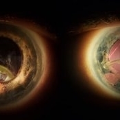
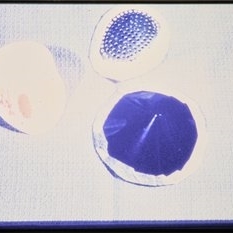 Eye Shields - Collection
-
 Giant Retinal Tear
Feb 20 2024 by Soobien Lee
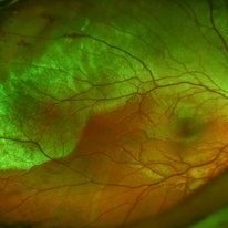
Optos color fundus photograph of a 40-year-old caucasian male who is a UFC fighter with a total retinal detachment in his right eye secondary to a giant retinal tear from 10 o'clock to 2 o'clock.
Photographer: Trinity Wolf, Elman Retina Group
Imaging device: Optos Ultra-Widefield Imaging
Condition/keywords: giant retinal tear, optos, Retinal Detachment, Retinal tear with detachment, trauma
-
 Giant Tear
Oct 28 2024 by Andreas Paulo Di Luciano Rojas, MD
Giant retinal tear secondary to trauma.
Photographer: Andreas Di-Luciano, MD
Imaging device: Optos
Condition/keywords: giant retinal tear, ocular trauma, proliferative vitreoretinopathy (PVR), retinectomy, Trauma
-
 Intraocular Foreign Body
Nov 8 2022 by pedro fernandes souza neto
Intraocular foreign body after stone trauma. CT scan image showing intraocular foreign body location.
Photographer: Pedro Fernandes, Federal University of Bahia
Condition/keywords: intraocular foreign body, trauma
-
 Intraocular Foreign Body
Feb 7 2019 by Somnath Chakraborty, MD
Left eye fundus photo montage of a 45-year-old male showing a large iron foreign body, impacted inferior to the infero-temporal branch vessels with a large patch of surrounding chorio-retinal atrophy, secondary to resolving Commotio retinae
Photographer: Saptarshi Mehta
Condition/keywords: commotio retinae, intraocular foreign body, trauma
-
 Intraocular Foreign Body
Mar 27 2019 by Gary R. Cook, MD, FACS
20-year-old white male who sustained penetrating ocular injury and vitreous hemorrhage OS from a piece of steel while hammering metal on metal; V.A.= 20/20-2.
Imaging device: Topcon VT-50
Condition/keywords: intraocular foreign body, non metallic retained intraocular foreign body (RIOFB), trauma, vitreous hemorrhage
-
 Intraocular Foreign Body with Retinal Detachment
Mar 27 2019 by Gary R. Cook, MD, FACS
49-year-old Asian male who sustained a penetrating ocular injury OD and a rhegmatogenous retinal detachment from this piece of metal; V.A.= 20/200.
Imaging device: Topcon VT-50
Condition/keywords: intraocular foreign body, trauma
 Loading…