 Initializing download.
-
 By Xitlali Caterina
Co-author(s): Gregory Bever, MD. Retina Specialist of Michigan - Uploaded on Mar 26, 2024.
- Last modified by Joshua Friedman on Mar 27, 2024.
- Rating
- Appears in
- Miscellaneous
- Condition/keywords
- Optos, OPTOS CALIFORNIA, ultra-wide field imaging, fundus photography, superior retina, ultra-widefield image
- Photographer
- Xitlali Caterina
- Imaging device
-
Scanning laser ophthalmoscope
Optos California RGB - Description
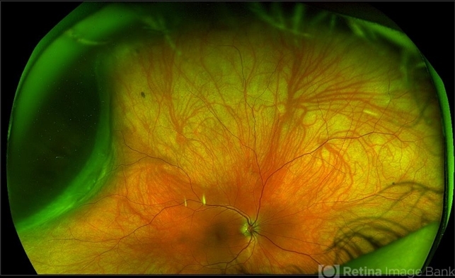
- Ultra-widefield fundus photograph of a 40-year-old woman with Choroidal Melanoma in right eye. Patient present with 20/50+2 vision in the right eye. Patient reported having frequent headaches located frontal area of their head and sometimes radiated to the right side as well. Patient also noted eye pain in both eyes that has remained constant for many years, as well as light sensitivity. The physician stated that since this is a medium-sized tumor, the treatment options include I-125 brachytherapy or enucleation. He recommended I-125 brachytherapy.


![Choroidal Nevus[004]](/Content/imagebank/Choroidal-Nevus-004---thumb.jpg/image-square;max$79,0.ImageHandler "Choroidal Nevus[004]")


