Search results (148 results)
-
 Vitreous Cavity Inhabitant
Jun 2 2025 by Poornachandra B, MS, FVRS
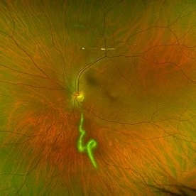
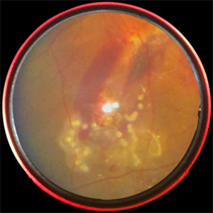
A 36-year-old male presented with a 6-week history of intermittent ocular redness, now accompanied by the recent onset of floaters for the past 2 days. Fundus examination revealed the presence of a nematode in the vitreous cavity.
Photographer: Mr Dhikshith
Condition/keywords: parasite
-
 Bot Fly Larvae
Apr 29 2025 by Daniela Bogenschutz
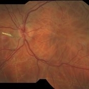
57 year-old male referred for decreased vision from optometrist. His only complaint was floaters and the letters were moving on the screen. He had never been out of the country, but is a farmer. Upon examination, our retina specialist found a bot fly larvae with numerous tracks made in this patient's retina. Patient was treated with laser to kill the larvae which was successful and he has been monitored yearly.
Photographer: Daniela Bogenschutz, OSC; Retina Consultants of Carolina, P.A.
Imaging device: Topcon
Condition/keywords: Bot Fly Larvae
-
 Choroidal Detachment
Jan 17 2022 by Logan ryzenga
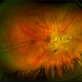
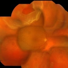
Left ultra-wide field photograph of an 81-year old female with a choroidal detachment affecting her left eye. Patient had a stent placed November, 2021 and following the procedure she complains of variable blurred vision and severe constricted visual fields. She presented at our office with flashes a month prior but without pain or floaters.
Photographer: Logan Ryzenga
Imaging device: Optos California
Condition/keywords: choroidal detachment, fundus photograph, left eye, Optos, pseudocolor, superior retina, ultra-wide field imaging
-
 Prominent Long Ciliary Nerve
Jan 25 2022 by Kachelle Brown
Ultra-wide field photograph of a 48-year-old female with a prominent long ciliary nerve. Patient presented asymptomatic, and was referred for a macula on retinal detachment. Patient was diagnosed with high myopia and a posterior vitreous detachment, and the physician discussed increased risk of floaters, myopic degeneration and retinal detachment associated with high myopia. -24.00 prior to cataract surgery OU per patient.
Photographer: Kachelle Brown
Imaging device: Optos California
Condition/keywords: fundus photograph, high myopia, long ciliary nerve, optos, right eye, ultra-widefield image
-
 Coats' Disease
Jul 16 2019 by Kim Barrett
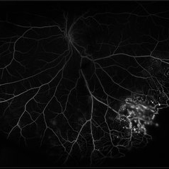
Ultra-wide field fluorescein angiogram of a 23-year-old male with Coats' disease, presented with distorted vision affecting his left eye. He reported seeing flashes and floaters since January of 2019, but the flashes had resolved. He was treated with Intravitreal Preservative Free Triamcinolone in the office and scheduled for PRP laser in the near future.
Photographer: Kim Barrett
Imaging device: Optos
Condition/keywords: Coats' disease, fluorescein angiogram (FA), fluorescein leakage, inferior retina, ischemia, left eye, Optos, ultra-wide field imaging
-
 Dislocated Capsular Tension Ring in Vitreous Cavity
Dec 21 2019 by Pablo Baquero Ospina, MD
Fundus photograph of an 52-year-old woman with pseudoexfoliation glaucoma and previous cataract surgery with capsular tension ring. 5 years later she refers floaters.
Photographer: Pablo Baquero, Asociacion Para Evitar la Ceguera en Mexico, Mexico city
Imaging device: Optos/Daytona
Condition/keywords: fundus photograph, pseudoexfoliation glaucoma
-
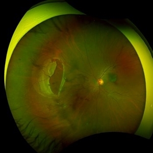
 Giant Retinal Tear
Jan 8 2017 by Manish Nagpal, MD, FRCS (UK), FASRS
Fundus photo of a patient complaining of floaters and a curtain like field defect.
Photographer: pravin jain
Condition/keywords: giant retinal tear
-
 Giant Retinal Tear Slide 1
Oct 22 2012 by Ronald C. Gentile, MD
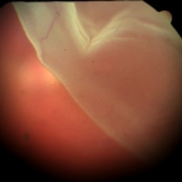
Acute loss of vision in a myopic man with flashes and floaters in the right eye. The giant retinal tear is flapped over with the macula detached. The undersurface of the retina can be seen temporally.
Photographer: The New York Eye & Ear Infirmary Department of Medical Imaging
Condition/keywords: retinal tear, vitrectomy
-
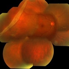
 Large Retinal Tear from a Shuttlecock Injury
Oct 11 2024 by Ahmad B. Tarabishy, MD
27 year old woman presenting with floaters and a shadow in her temporal visual field OS. Approximately one week earlier, she was injured in her left eye by a shuttlecock while playing badminton. Fundus exam reveals mild vitreous hemorrhage and a large retinal tear with a small cuff of surrounding SRF.
Photographer: Angela Rico, M.D.
Imaging device: Optos
Condition/keywords: blunt trauma, ocular trauma, retinal tear
-
 Methotrexate Bubble following Intravitreal Injection for PVR
Sep 21 2022 by Zach Seim
Ultra-widefield fundus photograph of an 81 year old female with a Methotrexate bubble following an Intravitreal Injection for Proliferative Vitreoretinopathy. Patient has been presenting to the office for two week interval Methotrexate injections in her left eye. The image was taken prior to her eighth injection which revealed a residual Methotrexate bubble in her inferior retinal image. This patient was seeing "lots" of floaters, as well as having visual acuity of cc20/400 cc20/200 PH.
Photographer: Zach Seim
Imaging device: OPTOS California
Condition/keywords: bubble, fundus photograph, fundus photography, intravitreal injection, left eye, methotrexate, nasal retina, Optos, proliferative vitreoretinopathy (PVR), pseudocolor, ultra-wide field imaging
-
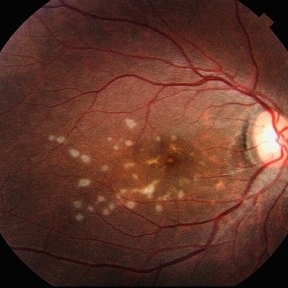 Multiple evanescent White Dot Syndrome (MEWDS)
May 27 2025 by César Adrián Gómez Valdivia, MD
Fundus photograph of a 21 year-old female patient with suspected Multiple Evanescent White Dot Syndrome (MEWDS). The White Dot Syndromes produce yellow-white retinal lesions classically located at the retinal pigment epithelium or outer retina and are found primarily in young adults. Symptoms of MEWDS include unilateral blurred vision, visual field loss, photopsias, and floaters.
Photographer: @eyemissu2
Imaging device: TOPCON TRC-50DX
Condition/keywords: multiple evanescent white dot syndrome (MEWDS)
-
 Posterior Scleral Laceration
May 24 2022 by Ahmad B. Tarabishy, MD
A 49 year old male was referred from the ER following an injury to his right medial eyelid with a sharp metal tip. He had brief pain at the time. No new floaters, flashes, or blurred vision. Intraocular pressure was 18 OS. Examination showed a full thickness laceration of the nasal posterior globe with adjacent hemorrhage. Prophylactic laser coagulation was performed. Examination 2 weeks later shows maturing laser scars and no complications related to the scleral laceration. The patient reports no new vision changes.
Photographer: Angelo Rico MD, Retina Specialists of Tampa
Imaging device: Optos
Condition/keywords: globe perforation, scleral laceration
-
 Retinal Detachment with Retinal Hole
Sep 30 2013 by Jason S. Calhoun
Patient in with complaints of floaters in the right eye. VA was 20/40 with no improvement. Fundus exam shows retinal detachment from 9-12 o'clock with hole at 10:30 posteriorly. Pneumatic retinopexy was performed with C3F8 Gas bubble and laser around the retinal tear in the right eye.
Photographer: Jason S. Calhoun, Department of Ophthalmology, Mayo Clinic Jacksonville, Florida
Imaging device: TOPCON TRC 50-EX
Condition/keywords: retinal hole
-
 Subretinal Worm with Laser Marks - Smartphone Fundus Photograph
Jun 13 2019 by Prithvi Chandrakanth
42-year-old, male came with chief complaints of diminished vision and floaters in right eye for past one week. On fundus examination noted to have subretinal haemorrhage and edema at the posterior pole and a subretinal live mobile worm at the periphery. Laser photocoagulation done followed by pars plana vitrectomy.
Photographer: Dr.PRITHVI CHANDRAKANTH, Dr.CHANDRAKANTH MALABAR NETHRALAYA, KOZHIKODE
Imaging device: TRASH TO TREASURE RETCAM - SMARTPHONE FUNDUS CAMERA DEVICE
Condition/keywords: laser photocoagulation, smartphone fundus photography, subretinal hemorrhage, uveitis, worm
-
 Vitreomacular Traction
Jun 15 2022 by Zach Seim
Optical Coherence Tomography (OCT) of a 69 year old male with Vitreomacular Traction affecting his right eye. Patient was referred to this office for Choroidal Melanoma in his right eye in May 2021. The patient was treated with Brachytherapy in July 2021 and this OCT was taken at a follow-up appointment in May 2022. Patient's vision was 20/30-2 at the time this OCT was taken. Patient states that his vision was better since his last visit, and that he sees floaters occasionally.
Photographer: Zach Seim
Imaging device: Heidelberg Spectralis
Condition/keywords: heidelberg spectralis, OD, optical coherence tomography (OCT), right eye, subretinal fluid, vitreomacular adhesion, vitreomacular interface disorders, vitreomacular traction (VMT)
-
 Rhegmatogenous Retinal Detachment
Aug 23 2012 by Gabriela Lopezcarasa Hernandez, MD
30-year-old male with floaters and inferonasal scotoma.
Photographer: Gabriela Lopezcarasa Hernandez, Hospital Angeles Lomas
Imaging device: ZEISS FF4
Condition/keywords: floaters, inferonasal scotoma
-
 Asteroid Hyalosis
Aug 27 2015 by René Hernán Parada Vásquez
Fundus photograph of 60-year-old male with an asteroid hyalosis, showing a multiple-yellow-white, round, particles composed of calcium.
Photographer: Parada René, ESO, Guatemala.
Condition/keywords: asteroid hyalosis, floaters
-
 Asteroid Hyalosis
Aug 23 2012 by Gabriela Lopezcarasa Hernandez, MD
56-year-old woman with floaters.
Photographer: Gabriela Lopezcarasa Hernandez, Hospital Angeles Lomas
Imaging device: ZEISS FF4
Condition/keywords: asteroid hyalosis, floaters
-
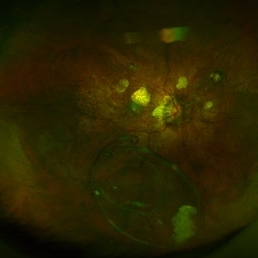
 Laser Barrage for Temporal Localized Rhematogenous Retinal Detachment
Feb 15 2018 by Kushal S Delhiwala, MBBS, MS, FMRF,FICO, FAICO
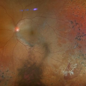
39-year-old female presenting with sudden onset flashes and floaters in left eye having undergone refractive surgery 20 years before for pathologic myopia.Color fundus photograph montage of left eye showing macula sparing inferotemporal localized Rhematogenous retinal detachment with horse shoe tear and temporal lattice degeneration treated with laser barrage.
Photographer: Dr Kushal Delhiwala, Netralaya superspeciality eye hospital ,Ahmedabad
Imaging device: Zeiss Visucam 500
Condition/keywords: barrier laser, macula sparring
-
 Subclinical RD with a Superior Tear
Feb 7 2018 by Manish Nagpal, MD, FRCS (UK), FASRS
Patient having 6/6 vision complaining of recent floaters and inferior field defect had a superior tear with surrounding retinal detachment.
Photographer: Mehul Prajapati
-
 GRT Grade B PVR
Dec 14 2018 by John S. King, MD
66-year-old white male with 6-8 weeks of decreased central vision OS following an episode of flashes and floaters, who presented to his eye doctor for cataract evaluation. Grade B PVR giant retinal tear / retinal detachment involving the macula. 20/200, RAPD, IOP 14
Photographer: Kay Dalby
Imaging device: Topcon 50
Condition/keywords: giant retinal tear, proliferative vitreoretinopathy (PVR)
-
 Actively Bleeding NVE
Apr 1 2025 by Jordyn Beckman
47 year old woman presented with actively bleeding NVE temporally on exam with complaints of foggy vision and floaters.
Photographer: Jordyn Beckman, Retina Consultants of Carolina, P.A.
Imaging device: Optos California
Condition/keywords: active bleeding, Elevated retinal neovascularization, vitreous hemorrhage
-
 ARN (#1) Initial Photo
May 27 2019 by John S. King, MD
60-year-old African American female who had been treated for iridocyclitis for at least a week sent in for vitritis and a nasal fundus lesion. Complaints included redness, floaters, photophobia, and decreased vision. Husband had recent shingles. Acuity was 20/60-2 with IOP of 12, and small KP in Art's triangel, 1-2+ a/c cell, 2-3+ ant vit cell, diffuse arteriolar sheathing, multiple areas of retinal whitening in periphery and mid-periphery (see Photo #1). PCR of a/c was performed, and intravitreal GCV administered, and VACV 2g qid and ASA started.... PCR positive for HZV, pred taper was started two days after presentation as the infection had begun to stablize..... Five days from presentation the vision was 20/60, inflammation and areas of retinal whitening had improved (see Photo #2).... One week later acuity was 20/30, the a/c was quiet and KP resolved; ant vitreous cell decreased; and there was further improvement in retinal appearance without any signs of retinal holes or detachment; she is now on low dose maint VACV (see photo#3)
Photographer: Maysee Yang
Imaging device: Optos CA
Condition/keywords: acute retinal necrosis, Herpes zoster
-
 ARN (#2) Five Days Since Initial Visit
May 27 2019 by John S. King, MD
60-year-old African American female who had been treated for iridocyclitis for at least a week sent in for vitritis and a nasal fundus lesion. Complaints included redness, floaters, photophobia, and decreased vision. Husband had recent shingles. Acuity was 20/60-2 with IOP of 12, and small KP in Art's triangel, 1-2+ a/c cell, 2-3+ ant vit cell, diffuse arteriolar sheathing, multiple areas of retinal whitening in periphery and mid-periphery (see Photo #1). PCR of a/c was performed, and intravitreal GCV administered, and VACV 2g qid and ASA started.... PCR positive for HZV, pred taper was started two days after presentation as the infection had begun to stablize..... Five days from presentation the vision was 20/60, inflammation and areas of retinal whitening had improved (see Photo #2).... One week later acuity was 20/30, the a/c was quiet and KP resolved; ant vitreous cell decreased; and there was further improvement in retinal appearance without any signs of retinal holes or detachment; she is now on low dose maint VACV (see photo#3)
Photographer: Maysee Yang
Imaging device: Optos CA
Condition/keywords: acute retinal necrosis, Herpes zoster
-
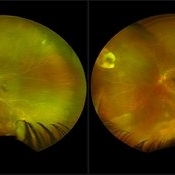 ARN (#3) This is comparison between the latest visit (left) and one week prior (which is the right photo, and same one as photo #2)
May 27 2019 by John S. King, MD
60-year-old African American female who had been treated for iridocyclitis for at least a week sent in for vitritis and a nasal fundus lesion. Complaints included redness, floaters, photophobia, and decreased vision. Husband had recent shingles. Acuity was 20/60-2 with IOP of 12, and small KP in Art's triangel, 1-2+ a/c cell, 2-3+ ant vit cell, diffuse arteriolar sheathing, multiple areas of retinal whitening in periphery and mid-periphery (see Photo #1). PCR of a/c was performed, and intravitreal GCV administered, and VACV 2g qid and ASA started.... PCR positive for HZV, pred taper was started two days after presentation as the infection had begun to stablize..... Five days from presentation the vision was 20/60, inflammation and areas of retinal whitening had improved (see Photo #2).... One week later acuity was 20/30, the a/c was quiet and KP resolved; ant vitreous cell decreased; and there was further improvement in retinal appearance without any signs of retinal holes or detachment; she is now on low dose maint VACV (see photo#3)
Photographer: Maysee Yang
Imaging device: Optos CA
Condition/keywords: acute retinal necrosis, Herpes zoster
 Loading…