Search results (7 results)
-
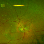
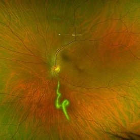 Vitreous Cavity Inhabitant
Jun 2 2025 by Poornachandra B, MS, FVRS
A 36-year-old male presented with a 6-week history of intermittent ocular redness, now accompanied by the recent onset of floaters for the past 2 days. Fundus examination revealed the presence of a nematode in the vitreous cavity.
Photographer: Mr Dhikshith
Condition/keywords: parasite
-
 Large Retinal Tear from a Shuttlecock Injury
Oct 11 2024 by Ahmad B. Tarabishy, MD
27 year old woman presenting with floaters and a shadow in her temporal visual field OS. Approximately one week earlier, she was injured in her left eye by a shuttlecock while playing badminton. Fundus exam reveals mild vitreous hemorrhage and a large retinal tear with a small cuff of surrounding SRF.
Photographer: Angela Rico, M.D.
Imaging device: Optos
Condition/keywords: blunt trauma, ocular trauma, retinal tear
-
 Methotrexate Bubble following Intravitreal Injection for PVR
Sep 21 2022 by Zach Seim
Ultra-widefield fundus photograph of an 81 year old female with a Methotrexate bubble following an Intravitreal Injection for Proliferative Vitreoretinopathy. Patient has been presenting to the office for two week interval Methotrexate injections in her left eye. The image was taken prior to her eighth injection which revealed a residual Methotrexate bubble in her inferior retinal image. This patient was seeing "lots" of floaters, as well as having visual acuity of cc20/400 cc20/200 PH.
Photographer: Zach Seim
Imaging device: OPTOS California
Condition/keywords: bubble, fundus photograph, fundus photography, intravitreal injection, left eye, methotrexate, nasal retina, Optos, proliferative vitreoretinopathy (PVR), pseudocolor, ultra-wide field imaging
-
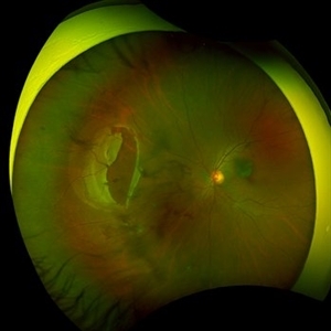
 Choroidal Detachment
Jan 17 2022 by Logan ryzenga
Left ultra-wide field photograph of an 81-year old female with a choroidal detachment affecting her left eye. Patient had a stent placed November, 2021 and following the procedure she complains of variable blurred vision and severe constricted visual fields. She presented at our office with flashes a month prior but without pain or floaters.
Photographer: Logan Ryzenga
Imaging device: Optos California
Condition/keywords: choroidal detachment, fundus photograph, left eye, Optos, pseudocolor, superior retina, ultra-wide field imaging
-
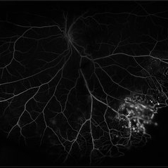 Coats' Disease
Jul 16 2019 by Kim Barrett
Ultra-wide field fluorescein angiogram of a 23-year-old male with Coats' disease, presented with distorted vision affecting his left eye. He reported seeing flashes and floaters since January of 2019, but the flashes had resolved. He was treated with Intravitreal Preservative Free Triamcinolone in the office and scheduled for PRP laser in the near future.
Photographer: Kim Barrett
Imaging device: Optos
Condition/keywords: Coats' disease, fluorescein angiogram (FA), fluorescein leakage, inferior retina, ischemia, left eye, Optos, ultra-wide field imaging
-
 Silicone Oil Large and Small Droplets 8 Days Post Avastin (Avella) Injection
Aug 16 2016 by Paul E. Tornambe, MD
The 88-year-old man had an Avastin injection (compounded by Avella) OD for AMD 8 days prior to this photo. He immediately complained of floaters after the injection which persisted 8 days later. The Optos photo, taken 8 days after the injection, shows a large silicone oil bubble about 1DD in size above the ST arcade and more than a dozen smaller 0.1DD bubbles over the macula suspended in the vitreous gel.
Photographer: Louanna Boren, Retina Consultants, San Diego
Condition/keywords: silicone oil, wet age-related macular degeneration (wet AMD)
-
 Rhegmatogenous Retinal Detachment
Aug 23 2012 by Gabriela Lopezcarasa Hernandez, MD
30-year-old male with floaters and inferonasal scotoma.
Photographer: Gabriela Lopezcarasa Hernandez, Hospital Angeles Lomas
Imaging device: ZEISS FF4
Condition/keywords: floaters, inferonasal scotoma
 Loading…