Search results (148 results)
-
---thumb.JPG/image-square;max$300,300.ImageHandler) Weiss Ring (Floater)
Weiss Ring (Floater)
Jul 10 2013 by Jason S. Calhoun
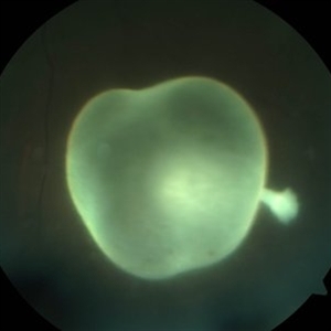
Patient comes in complaining of a floater towards the nasal aspect of her vision. Fundus photograph with anterior shot, shows a weiss ring pulled off from the optic nerve.
Photographer: Jason S. Calhoun, Department of Ophthalmology, Mayo Clinic Jacksonville, Florida
Condition/keywords: floaters, Weiss ring
-
 Asteroid Hyalosis
Aug 27 2015 by René Hernán Parada Vásquez
Fundus photograph of 60-year-old male with an asteroid hyalosis, showing a multiple-yellow-white, round, particles composed of calcium.
Photographer: Parada René, ESO, Guatemala.
Condition/keywords: asteroid hyalosis, floaters
-
---thumb.JPG/image-square;max$300,300.ImageHandler) Weiss Ring (Floater)
Weiss Ring (Floater)
Jul 10 2013 by Jason S. Calhoun
Patient comes in complaining of a floater towards the nasal aspect of her vision. Fundus photograph with anterior shot, shows a weiss ring pulled off from the optic nerve.
Photographer: Jason S. Calhoun, Department of Ophthalmology, Mayo Clinic Jacksonville, Florida
Condition/keywords: floaters, Weiss ring
-
 Asteroid Hyalosis
Aug 23 2012 by Gabriela Lopezcarasa Hernandez, MD
56-year-old woman with floaters.
Photographer: Gabriela Lopezcarasa Hernandez, Hospital Angeles Lomas
Imaging device: ZEISS FF4
Condition/keywords: asteroid hyalosis, floaters
-
 Rhegmatogenous Retinal Detachment
Aug 23 2012 by Gabriela Lopezcarasa Hernandez, MD
30-year-old male with floaters and inferonasal scotoma.
Photographer: Gabriela Lopezcarasa Hernandez, Hospital Angeles Lomas
Imaging device: ZEISS FF4
Condition/keywords: floaters, inferonasal scotoma
-
 Giant Retinal Tear Slide 1
Oct 22 2012 by Ronald C. Gentile, MD
Acute loss of vision in a myopic man with flashes and floaters in the right eye. The giant retinal tear is flapped over with the macula detached. The undersurface of the retina can be seen temporally.
Photographer: The New York Eye & Ear Infirmary Department of Medical Imaging
Condition/keywords: retinal tear, vitrectomy
-
 Toxoplasmosis Slide 1
Oct 22 2012 by Ronald C. Gentile, MD
Focal, white area of chorioretinitis with overlying vitreous inflammation adjacent to an old chorioretinal scar in a patient complaining of photophobia, floaters and a decrease in vision of the right eye. The focal area of chorioretinitis is involving the inferior nasal macula and adjacent optic nerve with surrounding retinal and peri-papillary edema.
Photographer: The New York Eye & Ear Infirmary Department of Medical Imaging
Condition/keywords: posterior uveitis, toxoplasmosis
-
---thumb.jpg/image-square;max$300,300.ImageHandler) Floaters
Floaters
Oct 9 2013 by Maurice F. Rabb
KR is a 25 year old white female who presented with a one month history of floaters OD. Past ocular and systemic history were unremarkable. On clinical examination, the visual acuity was 20/20 OU, and the anterior segments were normal. There was a very mild degree of vitreous cell OD, though no cystoid macular edema nor vasculitis. A lobulated white mass was noted overlying the vitreous base inferotemporally OD (thickness 3.3mm). There was no calcification, though prominent cysts were noted on the surface of the lesion. A fluorescein angiogram, echogram, and CT scan were obtained, along with a thorough systemic evaluation.
Condition/keywords: floaters
-
 Attached Vitreous With Floaters
Dec 10 2012 by Yale L. Fisher, MD
The vitreous is attached and demonstrates after-movements of formed vitreous as the patient is asked to look to the right and left. There is mild reflectivity in the formed vitreous from collagen. The optic nerve is visible in the superior aspect of the image and the lateral rectus muscle is seen inferiorly.
Condition/keywords: floaters, video, vitreous
-
---thumb.jpg/image-square;max$300,300.ImageHandler) Floaters And Blurred Vision
Floaters And Blurred Vision
Oct 21 2013 by Maurice F. Rabb
A 23 year old white female who presented with floaters and blurred vision in the left eye for 2 weeks.
Condition/keywords: blurred vision, floaters
-
 Paracentral Acute Middle Maculopathy (PAMM)
Mar 21 2019 by Jonathan C. Tsui, MD
26-year-old female with hypertension presenting with chief complaint of "darkening" in her nasal visual field in the right eye. No flashes, floaters, or vision loss. Va 20/60 and nasal VF defect OD. SD-OCT demonstrated hyperreflectivity in the INL consistent with paracentral acute middle maculopathy. She was referred to her PCP for blood pressure optimization and a cardiovascular work-up. She returned for follow-up two months later with 20/80 OD, 20/20 OS. Repeat SD-OCT demonstrated inner retinal atrophy.
Photographer: Zellers, Diane
Condition/keywords: paracentral acute middle maculopathy
-
---thumb.jpg/image-square;max$300,300.ImageHandler) Floaters
Floaters
Oct 9 2013 by Maurice F. Rabb
KR is a 25 year old white female who presented with a one month history of floaters OD. Past ocular and systemic history were unremarkable. On clinical examination, the visual acuity was 20/20 OU, and the anterior segments were normal. There was a very mild degree of vitreous cell OD, though no cystoid macular edema nor vasculitis. A lobulated white mass was noted overlying the vitreous base inferotemporally OD (thickness 3.3mm). There was no calcification, though prominent cysts were noted on the surface of the lesion. A fluorescein angiogram, echogram, and CT scan were obtained, along with a thorough systemic evaluation.
Condition/keywords: floaters
-
---thumb.jpg/image-square;max$300,300.ImageHandler) Floaters And Blurred Vision
Floaters And Blurred Vision
Oct 21 2013 by Maurice F. Rabb
A 23 year old white female who presented with floaters and blurred vision in the left eye for 2 weeks.
Condition/keywords: blurred vision, floaters
-
---thumb.jpg/image-square;max$300,300.ImageHandler) Floaters And Blurred Vision
Floaters And Blurred Vision
Oct 21 2013 by Maurice F. Rabb
A 23 year old white female who presented with floaters and blurred vision in the left eye for 2 weeks.
Condition/keywords: blurred vision, floaters
-
---thumb.jpg/image-square;max$300,300.ImageHandler) Floaters And Blurred Vision
Floaters And Blurred Vision
Oct 21 2013 by Maurice F. Rabb
A 23 year old white female who presented with floaters and blurred vision in the left eye for 2 weeks.
Condition/keywords: blurred vision, floaters
-
---thumb.jpg/image-square;max$300,300.ImageHandler) Floaters And Blurred Vision
Floaters And Blurred Vision
Oct 21 2013 by Maurice F. Rabb
A 23 year old white female who presented with floaters and blurred vision in the left eye for 2 weeks.
Condition/keywords: blurred vision, floaters
-
 Lasered Retinal Tear
Jul 14 2013 by Jason S. Calhoun
Patient with increased floaters. Fundus photos show retinal tear at 2-o'clock. Laser retinopexy was performed to prevent retinal detachment
Photographer: Jason S. Calhoun, Department of Ophthalmology, Mayo Clinic Jacksonville, Florida
Imaging device: TOPCON TRC 50-EX
Condition/keywords: laser retinopexy, laser treatment, retinal tear
-
 Laser Barrage for Temporal Localized Rhematogenous Retinal Detachment
Feb 15 2018 by Kushal S Delhiwala, MBBS, MS, FMRF,FICO, FAICO
39-year-old female presenting with sudden onset flashes and floaters in left eye having undergone refractive surgery 20 years before for pathologic myopia.Color fundus photograph montage of left eye showing macula sparing inferotemporal localized Rhematogenous retinal detachment with horse shoe tear and temporal lattice degeneration treated with laser barrage.
Photographer: Dr Kushal Delhiwala, Netralaya superspeciality eye hospital ,Ahmedabad
Imaging device: Zeiss Visucam 500
Condition/keywords: barrier laser, macula sparring
-
---thumb.JPG/image-square;max$300,300.ImageHandler) Giant Retinal Tear Treated With Laser
Giant Retinal Tear Treated With Laser
Jul 8 2013 by Jason S. Calhoun
Patient in with a shower of floaters. VA was 20/30 and fundus exam shows giant retinal tear temporally. Patient was treated with laser retinopexy to prevent retinal detachment.
Photographer: Jason S. Calhoun, Department of Ophthalmology, Mayo Clinic Jacksonville, Florida
Condition/keywords: laser retinopexy, retinal tear
-
---thumb.jpg/image-square;max$300,300.ImageHandler) Floaters And Blurred Vision
Floaters And Blurred Vision
Oct 21 2013 by Maurice F. Rabb
A 23 year old white female who presented with floaters and blurred vision in the left eye for 2 weeks.
Condition/keywords: blurred vision, floaters
-
 Retinal Detachment with Retinal Hole
Sep 30 2013 by Jason S. Calhoun
Patient in with complaints of floaters in the right eye. VA was 20/40 with no improvement. Fundus exam shows retinal detachment from 9-12 o'clock with hole at 10:30 posteriorly. Pneumatic retinopexy was performed with C3F8 Gas bubble and laser around the retinal tear in the right eye.
Photographer: Jason S. Calhoun, Department of Ophthalmology, Mayo Clinic Jacksonville, Florida
Imaging device: TOPCON TRC 50-EX
Condition/keywords: retinal hole
-
---thumb.jpg/image-square;max$300,300.ImageHandler) Floaters
Floaters
Oct 9 2013 by Maurice F. Rabb
KR is a 25 year old white female who presented with a one month history of floaters OD. Past ocular and systemic history were unremarkable. On clinical examination, the visual acuity was 20/20 OU, and the anterior segments were normal. There was a very mild degree of vitreous cell OD, though no cystoid macular edema nor vasculitis. A lobulated white mass was noted overlying the vitreous base inferotemporally OD (thickness 3.3mm). There was no calcification, though prominent cysts were noted on the surface of the lesion. A fluorescein angiogram, echogram, and CT scan were obtained, along with a thorough systemic evaluation.
Condition/keywords: floaters
-
 Intravitreal Cysticercosis
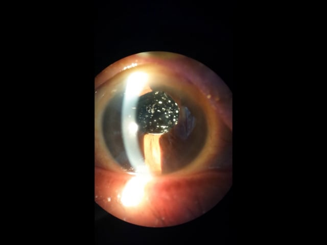
Apr 18 2014 by Neha Goel, MS DNB FRCS (Glasg)
A 35-year-old healthy male presented with complaints of floaters in his left eye since 2 weeks. On examination, visual acuity was 6/6, posterior pole was normal, the inferior fundus showed a live intravitreal cysticercus with a scolex, that was photophobic.
Photographer: Kiran Sharma, Guru Nanak Eye Centre, Maulana Azad Medical College, New Delhi, India
Imaging device: Zeiss Visucam
Condition/keywords: cysticercosis, intravitreal, scolex
-
---thumb.jpg/image-square;max$300,300.ImageHandler) Floaters
Floaters
Oct 9 2013 by Maurice F. Rabb
KR is a 25 year old white female who presented with a one month history of floaters OD. Past ocular and systemic history were unremarkable. On clinical examination, the visual acuity was 20/20 OU, and the anterior segments were normal. There was a very mild degree of vitreous cell OD, though no cystoid macular edema nor vasculitis. A lobulated white mass was noted overlying the vitreous base inferotemporally OD (thickness 3.3mm). There was no calcification, though prominent cysts were noted on the surface of the lesion. A fluorescein angiogram, echogram, and CT scan were obtained, along with a thorough systemic evaluation.
Condition/keywords: floaters
-
 Asteroid hyalosis
Aug 26 2015 by René Hernán Parada Vásquez
It is a video of a 60-year-old male with an asteroid hyalosis, this is a multiple-yellow-white, round, particles composed of calcium. You can see the movement of the vitreous when the patient looks up and down.
Photographer: Parada René, ESO, Guatemala
Condition/keywords: asteroid hyalosis, floaters
 Loading…