Search results (148 results)
-
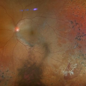
 Large Subhyaloid Hemorrhage
Jul 11 2025 by Jessilla Phou
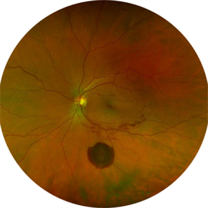
This is a fundus photograph depicting a large subhyaloid hemorrhage in the mid periphery of the left eye. The patient, a 53-year-old female, presented with a sudden onset of floaters, headache, and blurred vision. The image also demonstrates associated optic disc hemorrhage, vitreous hemorrhage, retinal hemorrhage, and venous tortuosity. Despite the extensive workup performed and the severity of the hemorrhage, no underlying cause was determined.
Photographer: Jessilla Phou
Imaging device: Optos California
Condition/keywords: fundus photograph, optic disc hemorrhage, retinal hemorrhage, venous tortuosity, vitreous hemorrhage
-
 Vitreous Cavity Inhabitant
Jun 2 2025 by Poornachandra B, MS, FVRS
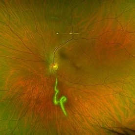
A 36-year-old male presented with a 6-week history of intermittent ocular redness, now accompanied by the recent onset of floaters for the past 2 days. Fundus examination revealed the presence of a nematode in the vitreous cavity.
Photographer: Mr Dhikshith
Condition/keywords: parasite
-
 Multiple evanescent White Dot Syndrome (MEWDS)
May 27 2025 by César Adrián Gómez Valdivia, MD
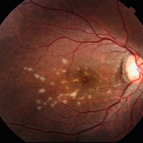
Fundus photograph of a 21 year-old female patient with suspected Multiple Evanescent White Dot Syndrome (MEWDS). The White Dot Syndromes produce yellow-white retinal lesions classically located at the retinal pigment epithelium or outer retina and are found primarily in young adults. Symptoms of MEWDS include unilateral blurred vision, visual field loss, photopsias, and floaters.
Photographer: @eyemissu2
Imaging device: TOPCON TRC-50DX
Condition/keywords: multiple evanescent white dot syndrome (MEWDS)
-
 Bot Fly Larvae
Apr 29 2025 by Daniela Bogenschutz
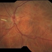
57 year-old male referred for decreased vision from optometrist. His only complaint was floaters and the letters were moving on the screen. He had never been out of the country, but is a farmer. Upon examination, our retina specialist found a bot fly larvae with numerous tracks made in this patient's retina. Patient was treated with laser to kill the larvae which was successful and he has been monitored yearly.
Photographer: Daniela Bogenschutz, OSC; Retina Consultants of Carolina, P.A.
Imaging device: Topcon
Condition/keywords: Bot Fly Larvae
-
 Actively Bleeding NVE
Apr 1 2025 by Jordyn Beckman
47 year old woman presented with actively bleeding NVE temporally on exam with complaints of foggy vision and floaters.
Photographer: Jordyn Beckman, Retina Consultants of Carolina, P.A.
Imaging device: Optos California
Condition/keywords: active bleeding, Elevated retinal neovascularization, vitreous hemorrhage
-
 Macular Degeneration
Dec 3 2024 by Sarah D Kang
Fundus photograph of an 85-year-old female patient with macular degeneration observed for retinal clearance before cataract surgery.
Condition/keywords: floaters, macular degeneration
-
 Macular Degeneration
Dec 3 2024 by Sarah D Kang
Fundus photograph of an 85-year-old female patient with macular degeneration observed for retinal clearance before cataract surgery.
Condition/keywords: floaters, macular degeneration
-
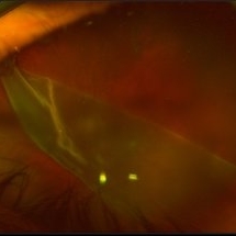
 Large Retinal Tear from a Shuttlecock Injury
Oct 11 2024 by Ahmad B. Tarabishy, MD
27 year old woman presenting with floaters and a shadow in her temporal visual field OS. Approximately one week earlier, she was injured in her left eye by a shuttlecock while playing badminton. Fundus exam reveals mild vitreous hemorrhage and a large retinal tear with a small cuff of surrounding SRF. This image was taken immediately following treatment with barrier laser retinopexy.
Photographer: Angela Rico, M.D.
Imaging device: Optos
Condition/keywords: blunt trauma, ocular trauma, retinal tear
-
 Large Retinal Tear from a Shuttlecock Injury
Oct 11 2024 by Ahmad B. Tarabishy, MD
27 year old woman presenting with floaters and a shadow in her temporal visual field OS. Approximately one week earlier, she was injured in her left eye by a shuttlecock while playing badminton. Fundus exam reveals mild vitreous hemorrhage and a large retinal tear with a small cuff of surrounding SRF.
Photographer: Angela Rico, M.D.
Imaging device: Optos
Condition/keywords: blunt trauma, ocular trauma, retinal tear
-
 Rhegmatogenous Macula Off Retinal Detachment with Multiple Breaks
May 29 2024 by Alexis Singstock
Ultra widefield fundus photograph of a 66 year old male with rhegmatogenous macula off retinal detachment with multiple breaks. Patient presented emergently for a curtain/veil in inferonasal visual field. Patient reports the curtain/veil in left eye started about 1 week prior, yet denied seeing flashes and floaters. Patient's vision was hand motion. Dr. Edward Korot examined the patient and scheduled him for a scleral buckle along with pars plana vitrectomy surgery.
Photographer: Alexis Singstock, Retina Specialists of Michigan
Imaging device: Optos California
Condition/keywords: fundus photography, left eye, macula off retinal detachment, OPTOS CALIFORNIA, pars plana vitrectomy (PPV), rhegmatogenous retinal detachment, scleral buckle, ULTRA WIDE FIELD
-
 Weiss Ring
Apr 22 2024 by SHIVANG CHAURASIA
Fundus photograph of a 68-year-old female with complaints of floaters.
Photographer: Dr SHIVANG CHAURASIA, GSVM MEDICAL COLLEGE, KANPUR, UTTAR PRADESH, INDIA
Imaging device: SMARTPHONE FUNDOSCOPY- IPHONE12
Condition/keywords: posterior vitreous detachment, Weiss ring
-
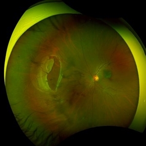
 Retinal Detachment with Giant Retinal Tear
Mar 26 2024 by Xitlali Caterina
Ultra-widefield fundus photograph of a 43-year-old male with a Retinal Detachment with Giant Retinal Tear affecting his left eye. Patient presented to the office with count fingers vision at 2 feet. He stated that about 8-9 days ago, he developed a clear curtain/veil and his vision started to get blurry. He also noted that he had floaters and flashes for about 8-9 days as well. The patient had cataract surgery a month prior to his visit. He stated that since his surgery, his vision had been better, but he had an area where he was not able to see well. The physician recommended a complex retinal detachment repair.
Photographer: Xitlali Caterina
Imaging device: OPTOS California RGB
Condition/keywords: fundus photograph, giant retinal tear, left eye, Optos, OPTOS CALIFORNIA, retinal detachment of the macula, retinal detachment with tear, ultra-wide field imaging, ultra-widefield image
-
 Branch Retinal Vein Occlusion with Retinal Neovascularization
Mar 21 2024 by Isaac Agranoff
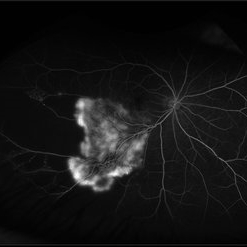
Fundus angiography photograph of a 63 year old male presenting with worsening blurry vision OD for 4 years with new transient floaters (vision 20/160 PH 20/60). Fluorescein angiography revealed significant capillary non-perfusion corresponding to the area, with peripheral vascular remodeling. Physician recommended anti-VEGF therapy and FA-guided supplemental PRP given the size of the NVE.
Photographer: Isaac Agranoff
Imaging device: Optos California
Condition/keywords: branch retinal vein occlusion (BRVO), EYLEA, FLUORESCEIN ANGIOGRAPHY, Neovascularisation elsewhere (NVE), Optos
-
 Roth Spots
Mar 5 2024 by James P Dossett, MD
Pseudocolor fundus photograph of the right eye of a 56-year-old man who presented for evaluation of floaters noted to have bilateral Roth spots on dilated fundus exam. WBC count was obtained and was >300k. Bone marrow biopsy was performed and was consistent with chronic myelogenous leukemia. He was started on dasatinib and hydroxycarbamide. 1 month later the hemorrhages had improved significantly.
Imaging device: Optos
Condition/keywords: Roth spots
-
 Asteroid Hyalosis
Mar 2 2024 by Boutayna Azarkan , Doctor
Fundus photograph of a 67 years old patient who presented with significant floaters.
Photographer: BOUTAYNA AZARKAN
Imaging device: optos
Condition/keywords: asteroid hyalosis
-
 Asteroid Hyalosis
Mar 2 2024 by Boutayna Azarkan , Doctor
Fundus photograph of a 67 years old patient who presented with significant floaters.
Photographer: BOUTAYNA AZARKAN
Imaging device: OPTOS
Condition/keywords: Asteroid Hyalosis
-
 Peripheral retinal degenerations
Jan 29 2024 by Anupama Kiran Kumar
Fundus photo of a young man who underwent barrage laser after he presented to the clinic with floaters and was diagnosed to have lattices with horse shoe tears and retinal holes.
Photographer: Dr Anupama Kiran Kumar DNB FVR , Narayana Nethralaya Bangalore
Imaging device: Mirante SLO/OCT (Nidek Co., Gamagori, Japan)
Condition/keywords: lattice degeneration, peripheral retinal degeneration
-
 Choroidal Melanoma
Sep 7 2023 by Annaka Gooding
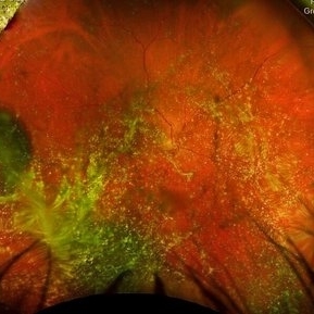
Ultra-Widefield pseudo-color and autofluorescence imaging of a 59 year old male with Choroidal Melanoma affecting his left eye. Patient reported floaters OS for months prior to examination as well as 1-2 weeks of "tunnel vision". Patient denies personal history of cancer. Patient's vision at time of examination was CF@5FT. Due to the Tumor size, the patient has developed a serous retina detachment in their inferior retina
Photographer: Annaka Gooding
Imaging device: Optos California
Condition/keywords: autofluorescence imaging, choroidal tumor, fundus photography, OPTOS CALIFORNIA, serous retinal detachment
-
 Retinal Detachment OD
Aug 23 2023 by Angela Rico
59 year-old male presents with Flashes and floaters OD for 2-3 weeks after Cataract surgery
Photographer: Angela Rico M.D.
-
 FA Malignant Melanoma
Aug 18 2023 by Angela Rico
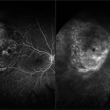
A 48 year old female who presented with peripheral flashes and floaters , 1 month duration Fluorescein angiogram of Malignant Melanoma. Picture on left- Timer 00:08:163 Picture on right- Timer 04:08:92
Photographer: Angela Rico M.D.
Condition/keywords: malignant melanoma
-
 Malignant Melanoma
Aug 18 2023 by Angela Rico
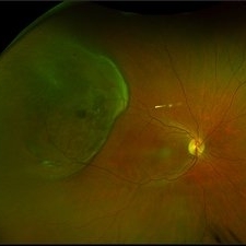
Fundus photograph of a 48 year old female with history of peripheral flashes and floaters OD for one month
Photographer: Angela Rico M.D.
Condition/keywords: intraocular tumor, malignant melanoma
-
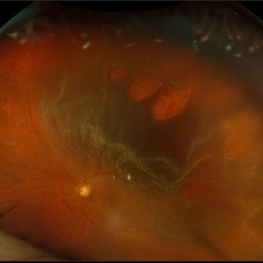
 Leukemic Retinopathy - OS
Aug 1 2023 by Shaleen Arora
A 14-year-old female was transferred from an outside hospital with a new diagnosis of B-ALL and WBC of 667,000. Following lumbar puncture, she developed blurry vision and floaters but denied curtaining, flashes, and diplopia. Ophthalmology was consulted to assess for disc edema. Exam revealed visual acuity of 20/100 OD and 20/200 OS. Imaging showed diffuse hemorrhages and Roth spots OU, consistent with leukemic retinopathy. The patient was followed by retinal specialists with spontaneous improvement in visual acuity over three weeks.
Photographer: Camilo Martinez, Childrens National Medical Center, Department of Ophthalmology
Condition/keywords: leukemia, leukemic infiltration, retinopathy, Roth spots
-
 Leukemic Retinopathy - OD
Aug 1 2023 by Shaleen Arora
A 14-year-old female was transferred from an outside hospital with a new diagnosis of B-ALL and WBC of 667,000. Following lumbar puncture, she developed blurry vision and floaters but denied curtaining, flashes, and diplopia. Ophthalmology was consulted to assess for disc edema. Exam revealed visual acuity of 20/100 OD and 20/200 OS. Imaging showed diffuse hemorrhages and Roth spots OU, consistent with leukemic retinopathy. The patient was followed by retinal specialists with spontaneous improvement in visual acuity over three weeks.
Photographer: Camilo Martinez, Childrens National Medical Center, Department of Ophthalmology
Condition/keywords: leukemia, leukemic infiltration, retinopathy, Roth spots
-
 Birdshot Retinopathy
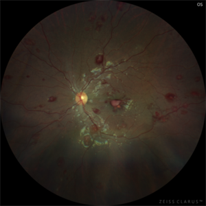
May 9 2023 by JEFFERSON R SOUSA, Tecg.º (Biomedical Systems Technology)
Female patient, 41 years old, with progressive low visual acuity, progressive history of autoimmune disease. In the multimodal retinal fundoscopic evaluation, important characteristics compatible with "Birdshot Retinopathy" were observed. Birdshot retinopathy, also known as birdshot chorioretinopathy or birdshot uveitis, is a rare, chronic inflammatory disorder that affects the retina and the choroid of the eye. It typically develops in adults between the ages of 30 and 60 years, and is more common in women than men. The name "birdshot" refers to the small, round, yellow-white spots that appear on the retina, which resemble the pattern of a shotgun blast. These spots are caused by inflammation in the eye, and can lead to vision loss if left untreated. Symptoms of birdshot retinopathy include blurred vision, floaters, loss of night vision, and difficulty adapting to changes in lighting. The condition can also cause inflammation in other parts of the eye, leading to redness, pain, and sensitivity to light. The exact cause of birdshot retinopathy is unknown, but it is believed to be an autoimmune disorder, in which the body's immune system mistakenly attacks the retina and choroid. Treatment typically involves the use of immunosuppressive medications, such as corticosteroids or biologic agents, to reduce inflammation and preserve vision. Close monitoring by an ophthalmologist is important, as the disease can progress even with.
Photographer: JEFFERSON ROCHA DE SOUSA - Retinal Department at Institute Dr. Suel Abujamra Sao Paulo-Brazil
Imaging device: Clarus 700 - Zeiss, composite of four 135 degree images.
Condition/keywords: bilateral chorioretinal folds, birdshot, birdshot chorioretinopathy, birdshot choroidopathy, birdshot retinochoroidopathy
-
 Birdshot Retinopathy
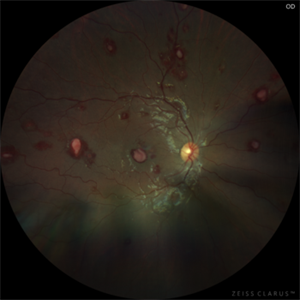
May 9 2023 by JEFFERSON R SOUSA, Tecg.º (Biomedical Systems Technology)
Female patient, 41 years old, with progressive low visual acuity, progressive history of autoimmune disease. In the multimodal retinal fundoscopic evaluation, important characteristics compatible with "Birdshot Retinopathy" were observed. Birdshot retinopathy, also known as birdshot chorioretinopathy or birdshot uveitis, is a rare, chronic inflammatory disorder that affects the retina and the choroid of the eye. It typically develops in adults between the ages of 30 and 60 years, and is more common in women than men. The name "birdshot" refers to the small, round, yellow-white spots that appear on the retina, which resemble the pattern of a shotgun blast. These spots are caused by inflammation in the eye, and can lead to vision loss if left untreated. Symptoms of birdshot retinopathy include blurred vision, floaters, loss of night vision, and difficulty adapting to changes in lighting. The condition can also cause inflammation in other parts of the eye, leading to redness, pain, and sensitivity to light. The exact cause of birdshot retinopathy is unknown, but it is believed to be an autoimmune disorder, in which the body's immune system mistakenly attacks the retina and choroid. Treatment typically involves the use of immunosuppressive medications, such as corticosteroids or biologic agents, to reduce inflammation and preserve vision. Close monitoring by an ophthalmologist is important, as the disease can progress even with.
Photographer: JEFFERSON ROCHA DE SOUSA - Retinal Department at Institute Dr. Suel Abujamra Sao Paulo-Brazil
Imaging device: Clarus 700 - Zeiss, composition of five 135 degree images.
Condition/keywords: bilateral chorioretinal folds, birdshot, birdshot chorioretinopathy, birdshot choroidopathy, birdshot retinochoroidopathy
 Loading…