Search results (959 results)
-
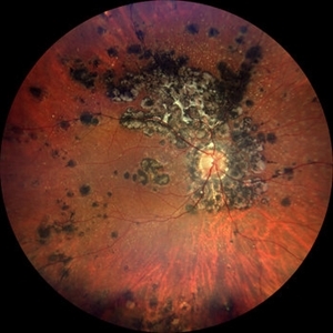 Tuberculosis-related serpiginous-like choroiditis
Nov 22 2022 by Ricardo Leitão Guerra
True color BLFI of a 60-year-old male presenting chorioretinal scars from a tuberculosis-related serpiginous-like choroiditis.
Photographer: Ricardo Leitão Guerra
Imaging device: Zeiss Clarus 700
Condition/keywords: serpiginous choroiditis, tuberculosis
-
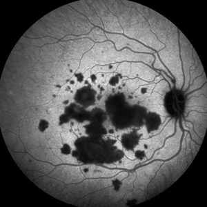 Relentless Placoid Chorioretinitis
Jan 9 2019 by Janet Brazil
ICG photo of a 24-year-old male with Relentless placoid chorioretinitis
Photographer: Janet Atkinson, Eye Associates of New Mexico
Imaging device: Heidelberg HRA Spectralis
Condition/keywords: placoid retinal lesions
-
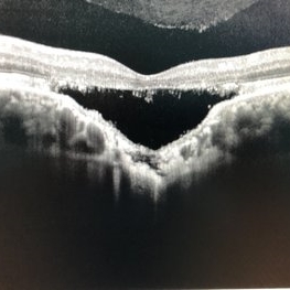 Choroidal Excavation
Jun 2 2019 by Nelson Chamma Capelanes, MD
SD-OCT of a 32-year-old woman showing a subfoveal choroidal excavation associated with chronic central serous chorioretinopathy.
Photographer: Nelson Chamma Capelanes, Promacula, Brazil
Imaging device: Heidelberg Spectralis SD-OCT
Condition/keywords: choroidal excavation, chronic central serous chorioretinopathy (CSCR), pachychoroid
-
 Pigmented Paravenous Retinochoroidal Atrophy (PPRCA)
Jun 30 2025 by Maria Letícia Costa Holanda
Fundoscopy of a 42-year-old asymptomatic man with pigmented paravenous chorioretinal atrophy. Pigmented paravenous retinochoroidal atrophy (PPRCA) is a rare disorder of unknown etiology. The disease is characterized by pigment accumulation along the distribution of retinal veins. The findings are usually incidental with minimal effect on vision.
Photographer: Guilherme da Cruz Reis, CLINOS Eye Hospital - Feira de Santana (BA),Brazil
Condition/keywords: pigmented paravenous chorioretinal atrophy (PPCRA)
-
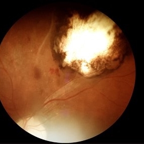
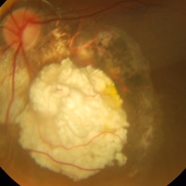 Retinoblastoma Regressed
Dec 31 2015 by P. Mahesh Shanmugam, MBBS, DO, FRCSEd, PhD, FAICO
Regressed Retinoblastoma S/P chemotherapy and multiple sessions of TTT. Central calcific residue with surrounding chorio-retinal atrophy is well noted.
Condition/keywords: retinoblastoma
-
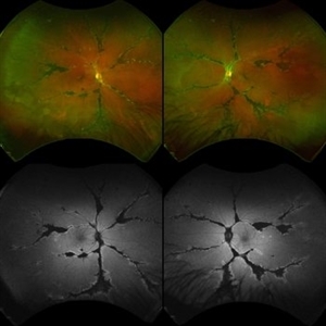
 Rod Cone dystrophy
Nov 29 2022 by Niloofar Piri, MD
Fundus photograph of the left eye in a 58 yo male with rod cone dystrophy. He presented with night blindness and peripheral vision loss since youth and recent decrease in central vision for the past 10 years. Notice waxy pallor of the nerve, severe arterial narrowing and chorioretinal atrophy mainly around the arcades as well as posterior pole along with RPE hyperplastic changes and atrophy. RPE atrophy in midperiphery has coin shaped appearance. FAF has characteristic appearance (uploaded separately) He has one pathogenic variants of both CEP290 and PRPH2 genes.
Photographer: Sean Kelso, Saint Louis University
Condition/keywords: hereditary retinal deg, hereditary retinal dystrophy, Rod cone dystrophy
-
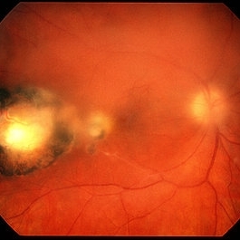
 Acute Syphilitic Posterior Placoid Chorioretinitis
Oct 16 2024 by César Adrián Gómez Valdivia, MD
Fundus autofluorescence image of an acute syphilitic posterior placoid chorioretinitis found in a HIV positive 28 YO male patient with suspected neurosyphilis. A beautiful butterfly autofluorescence pattern can be appreciated.
Photographer: @eyemissu2
Imaging device: California ICG OPTOS
Condition/keywords: acute syphilitic posterior placoid chorioretinitis, chorioretinitis, syphilis
-
 Acute Syphilitic Posterior Placoid Chorioretinitis
Sep 3 2016 by ADRIANO FERREIRA
66-year-old woman with acute visual acuity loss.
Photographer: Claudio Zett Lobo, UNIFESP
Condition/keywords: acute syphilitic posterior placoid chorioretinitis
-
 Cat Eye Syndrome
Feb 11 2020 by Sophia El Hamichi, MD
A 3-year-old female with cat eye syndrome including iris, chorioretinal and optic nerve colobomas. Note the CNV temporally to the optic nerve coloboma (blue arrows)
Photographer: Giselle De Oliveira, Bascom Palmer Eye Institute, Miami
Imaging device: RetCam
Condition/keywords: cat eye syndrome, chorioretinal coloboma, choroidal neovascularization (CNV), coloboma, coloboma of optic disc, optic nerve coloboma
-
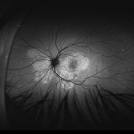
 Central Serous Retinopathy
Mar 19 2024 by Corey Grant
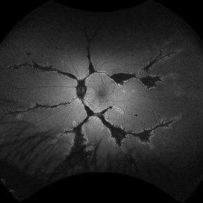
Ultra Wide-Field Fundus Autofluorescence Imaging of a 37 year old female with Central Serous Retinopathy affecting her right eye. Patient Visual Acuity was 20/20 in both eyes. Patient reported black spots in her vision onset three years ago, with associating flashes of light. Patient reports history of cortisone back injections a few years ago and denies Flonase use. The physician stated that there is hyperautofluorescence in the area of gutter of Sub-Retinal Fluid which likely happened from CSR.
Photographer: Corey Grant, OSC
Imaging device: OPTOS CALIFORNIA RGB
Condition/keywords: Central Serous Chorioretinopathy (CSR), central serous retinopathy (CSR), fundus autofluorescence (FAF), Guttering, hyperautofluorescence, inferior retina, OPTOS, Retina, Right Eye, subretinal fluid, ULTRA WIDE FIELD
-
 Chorioretinitis with Overlying Vitreous Stranding/Vitritis
Mar 23 2023 by Isaac Agranoff
Fundus photograph of a 37-year-old woman presenting with chorioretinitis with overlying vitreous stranding/vitritis that has remained unchanged for multiple years. Patient presented with irritation and blurred vision and her vision was 20/40 OD. The OCT revealed evidence of low-grade inflammation and the recommend treatment was anti-inflammatory eye drops at this time and to obtain second opinion with another physician in the office.
Photographer: Isaac Agranoff, Technician
Imaging device: Optos California
Condition/keywords: chorioretinal scar, chorioretinitis, inflammation, Optos, ultra-wide field imaging, vitritis
-
 Congenital Toxoplasmosis
Feb 2 2021 by Niloofar Piri, MD
43-year-old female with large oval chorioretinal scar in posterior pole with heavy RPE hyperplasia and history of hydrocephalus s/p VP shunt since birth. Findings are consistent with congenital toxoplasmosis.
Condition/keywords: congenital toxoplasmosis
-
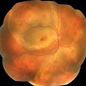
 Diffuse Chorioretinal Atrophy
Feb 21 2024 by Virginia Gebhart
61 year male with myopic degeneration and diffuse chorioretinal atrophy. BCVA 20/200.
Photographer: Virginia Gebhart
Imaging device: Topcon TRC 50DX
Condition/keywords: chorioretinal atrophy, myopic degeneration
-
 Idiopathic Uveal Effusion Syndrome
Aug 22 2024 by Jordyn Beckman
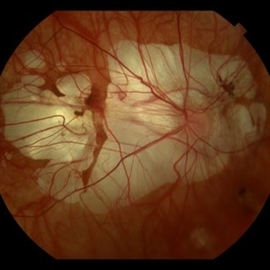
61 year old male with Idiopathic Uveal Effusion Syndrome with starry night appearance on fluorescein. 3 weeks s/p single external drainage retinotomy and 9 weeks of oral pred with recurrent choroidal effusions. Has since returned to surgery for secondary drainage retinotomy; subretinal fluid remain persistent.
Photographer: Jordyn Beckman
Imaging device: Optos California
Condition/keywords: chorioretinitis, Choroidal, exudative detachment, window defect
-
 Multifocal CSR With Exudative RD
May 19 2017 by Manish Nagpal, MD, FRCS (UK), FASRS
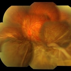
A 30-year-old male diagnosed elsewhere as VKH was started on heavy steroids and he developed multiple serous elevations and OS developed a exudative RD.
Photographer: POOJA BAROT
Condition/keywords: central serous retinopathy (CSR), multifocal central serous chorioretinopathy (CSCR), Vogt-Koyanagi-Harada
-
 PEHCR (Peripheral Exudative Hemorrhagic Chorioretinopathy)
May 12 2023 by Niloofar Piri, MD
Ultrawide fundus photograph of the left eye demonstrating extensive peripheral hemorrhagic exudative detachment in a 79 yo Caucasian female with prior history of non-exudative AMD. Recent diagnosis of Acute myeloid leukemia with low platelet count which might have contributed to the above presentatuon. Please note the temporal subretinal hemorrhage as well as RPE atrophy and hyperplasia in the macula.
Photographer: Rocio Bentivegna, MD, Saint Louis University; Jessica Maddox, COA, Saint Louis University
Condition/keywords: peripheral exudative hemorrhagic chorioretinopathy (PEHCR)
-
 Pigmented Paravenous Chorioretinal Atrophy (PPCRA)
Jun 27 2025 by Maria Letícia Costa Holanda
Fundoscopy of a 42-year-old asymptomatic man with pigmented paravenous chorioretinal atrophy. Pigmented paravenous retinochoroidal atrophy (PPRCA) is a rare disorder of unknown etiology. The disease is characterized by pigment accumulation along the distribution of retinal veins. The findings are usually incidental with minimal effect on vision.
Photographer: Guilherme da Cruz Reis, CLINOS Eye Hospital - Feira de Santana (BA),Brazil
Condition/keywords: pigmented paravenous chorioretinal atrophy (PPCRA)
-
 Pigmented Paravenous Retinochoroidal Atrophy (PPRCA)
Jun 27 2025 by Maria Letícia Costa Holanda
Fundoscopy of a 42-year-old asymptomatic man with pigmented paravenous chorioretinal atrophy. Pigmented paravenous retinochoroidal atrophy (PPRCA) is a rare disorder of unknown etiology. The disease is characterized by pigment accumulation along the distribution of retinal veins. The findings are usually incidental with minimal effect on vision.
Photographer: Guilherme da Cruz Reis, CLINOS Eye Hospital - Feira de Santana (BA),Brazil
Condition/keywords: pigmented paravenous chorioretinal atrophy (PPCRA)
-
 Retinocoroiditis Inactiva Por Toxoplasmosis
Apr 28 2025 by Paulina Araujo
Fundus photography demonstrates a 2-disc-diameter chorioretinal scar in the superior temporal arcade, consistent with inactive toxoplasmic retinochoroiditis. The lesion exhibits pigmented borders and central atrophy, with adjacent splinter hemorrhages and vascular sheathing. No vitreous inflammation or active satellite lesions are present.
Photographer: Paulina D.Araujo Martínez, Asociación para Evitar la Ceguera en México I.A.P., Hospital Dr Luis Sánchez Bulnes.
Condition/keywords: toxoplasmosis chorioretinitis
-
 Sunset Glow Fundus
May 15 2022 by Manuel Ángel Alcántara Delgado, MD
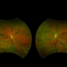
Optomap ultra-widefield retinal imaging of an 35-year-old woman showed sunset glow fundus, multiple nummular chorioretinal atrophic lesions, macular subretinal fibrosis and pigment clumping in chronic recurrent stage of Vogt-Koyanagi-Harada disease.
Photographer: Manuel Ángel Alcántara Delgado. Conde de Valenciana.
Condition/keywords: abnormal retina, benign pigmented lesions, pigment clumps, retinal fibrosis, uveitis, Vogt-Koyanagi-Harada
-
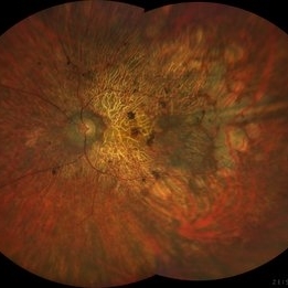
 Thioridazine-toxicity
Apr 30 2022 by Niloofar Piri, MD
61 yo male with PMH of longstanding schizophrenia since 20s with secondary intellectual disability presented with decreased vision following a recent stroke. He was found to have bilateral chorio-retinal atrophy involving posterior pole with scalloped edges and coin shaped atrophic area at margins extending into mid-periphery, diagnosis most concerning for intermediate stage thioridazine toxicity given the history. Mother could find handwritten prescriptions from 1990s when he was on Thioridazine 800 mg daily for unknown period of time. Patient had better vision in the left eye which was affected by recent stroke and prompted him to seek medical care. Fundus photograph of the right eye is demonstrated here.
Photographer: Jacob Grodsky, MD
Condition/keywords: drug toxicity, thioridazine toxicity, toxic retinopathy
-
 Toxo Lesion
Dec 29 2016 by Manish Nagpal, MD, FRCS (UK), FASRS
Healing toxo chorioretinitis lesion post a course of anti-toxo regime.
Photographer: Avijit Vishnoi
Condition/keywords: toxoplasmosis chorioretinitis
-
 Toxoplasma Retinochoroiditis
Feb 25 2013 by Henry J. Kaplan, MD
Toxoplasmosis, right eye: reactivation of congenital toxoplasmosis as an active retinitis lesion with overlying vitritis adjacent to an old scar.
Condition/keywords: toxoplasmosis chorioretinitis, toxoplasmosis reactivation
-
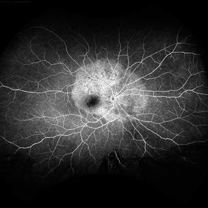 Acute Syphilitic Posterior Placoid Chorioretinitis
May 4 2021 by RAFAEL REIS PEREIRA, MD
A 31-year-old patient with a complaint of photophobia and low visual acuity OD in the previous three weeks. BCVA was 20/60 and 20/20 The fundus examination revealed a placoid white lesion in the posterior pole and vitreous cells in the right eye. The left eye was unremarkable. Fluorescein angiography reveals hyperfluorescent plaque with distinctive “leopard spots” hypofluorescence.
Imaging device: Opto California
Condition/keywords: acute syphilitic posterior placoid chorioretinitis
-
 Acute syphilitic posterior placoid chorioretinitis
Apr 24 2022 by Aniruddha K Agarwal, MD
Green-light fundus autofluorescence (FAF) of the right eye from a 55-year-old man with risk factors for sexually trasnmitted diseases who presented to the retina clinic for a central scotoma. Funduscopy revealed a placoid lesion in the posterior pole. FAF highlights a hyperautofluorescent placoid lesion involving the macula with granular hyperfluorescence. The patient tested positive for syphilis and received intravenous penicillin treatment.
Photographer: Esther CIANCAS, MD, PhD, Gema CRESPO-RODRÍGUEZ, RN
Imaging device: Zeiss Clarus fundus camera
Condition/keywords: chorioretinitis, IUSG, syphilis, uveitis
 Loading…