Search results (33 results)
-
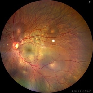
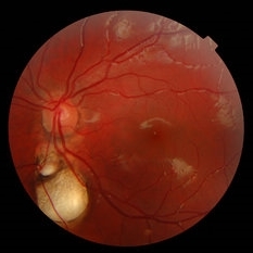
 Acute Syphilitic Posterior Placoid Chorioretinitis
Oct 20 2024 by César Adrián Gómez Valdivia, MD
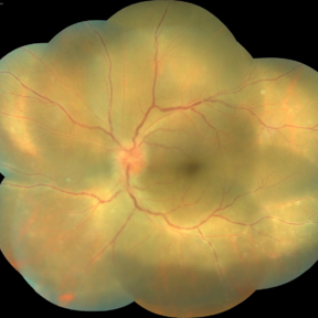
Fundus autofluorescence image of an acute syphilitic posterior placoid chorioretinitis found in a HIV positive 28 YO male patient with suspected neurosyphilis. A beautiful butterfly autofluorescence pattern can be appreciated.
Photographer: @eyemissu2
Imaging device: California ICG OPTOS
Condition/keywords: acute syphilitic posterior placoid chorioretinitis
-
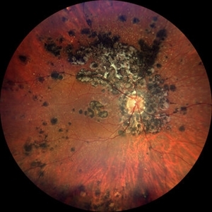
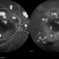
 Idiopathic Uveal Effusion Syndrome
Aug 22 2024 by Jordyn Beckman
61 year old male with Idiopathic Uveal Effusion Syndrome with starry night appearance on fluorescein. 3 weeks s/p single external drainage retinotomy and 9 weeks of oral pred with recurrent choroidal effusions. Has since returned to surgery for secondary drainage retinotomy; subretinal fluid remain persistent.
Photographer: Jordyn Beckman
Imaging device: Optos California
Condition/keywords: chorioretinitis, Choroidal, exudative detachment, window defect
-
 Central Serous Chorioretinopathy in Pregnancy (OS)
Apr 28 2024 by Vishal Agrawal, MD, FRCS,FACS,FASRS
30-year female with sudden loss of vision came for examination. She was in her first trimester of pregnancy. Examination revealed bilateral bullous NSD with subretinal fibrin s/o CSR.
Photographer: Dr Ayushi
Imaging device: Clarus 700
Condition/keywords: Central Serous Chorioretinopathy (CSR), neurosensory detachment of retina, pregnancy
-
 Chorioretinal Scar
Feb 19 2024 by Sanauddin Samejo , Diploma (Ophthalmic Technician Training Course)
A patient came in to the clinic of Dr Madhav Rao (VR Surgeon).
Photographer: Sanauddin Samejo, Burjeel Hospital, Abu Dhabi, UAE.
Imaging device: Optos Silverstone
Condition/keywords: retinal scar
-
 Gyrate Atrophy
Apr 12 2023 by Ahmed Abbas Hashmi, OD
Left eye fundus of a 53-year-old male patient with advanced gyrate atrophy of the choroid and retina with macular sparing. Optic nerve head is healthy.
Photographer: Ahmed Abbas Hashmi
Imaging device: Topcon TRC-NW8F
Condition/keywords: chorioretinal atrophy
-
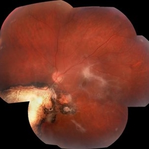
 PEHCR (Peripheral Exudative Hemorrhagic Chorioretinopathy)
May 12 2023 by Niloofar Piri, MD
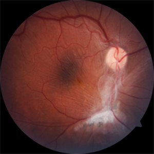
Ultrawide fundus photograph of the left eye demonstrating extensive peripheral hemorrhagic exudative detachment in a 79 yo Caucasian female with prior history of non-exudative AMD. Recent diagnosis of Acute myeloid leukemia with low platelet count which might have contributed to the above presentatuon. Please note the temporal subretinal hemorrhage as well as RPE atrophy and hyperplasia in the macula.
Photographer: Rocio Bentivegna, MD, Saint Louis University; Jessica Maddox, COA, Saint Louis University
Condition/keywords: peripheral exudative hemorrhagic chorioretinopathy (PEHCR)
-
 Tuberculosis-related serpiginous-like choroiditis
Nov 22 2022 by Ricardo Leitão Guerra
True color BLFI of a 60-year-old male presenting chorioretinal scars from a tuberculosis-related serpiginous-like choroiditis.
Photographer: Ricardo Leitão Guerra
Imaging device: Zeiss Clarus 700
Condition/keywords: serpiginous choroiditis, tuberculosis
-
 Thioridazine-toxicity
Apr 30 2022 by Niloofar Piri, MD
61 yo male with PMH of longstanding schizophrenia since 20s with secondary intellectual disability presented with decreased vision following a recent stroke. He was found to have bilateral chorio-retinal atrophy involving posterior pole with scalloped edges and coin shaped atrophic area at margins extending into mid-periphery, diagnosis most concerning for intermediate stage thioridazine toxicity given the history. Mother could find handwritten prescriptions from 1990s when he was on Thioridazine 800 mg daily for unknown period of time. Patient had better vision in the left eye which was affected by recent stroke and prompted him to seek medical care. Fundus photograph of the right eye is demonstrated here.
Photographer: Jacob Grodsky, MD
Condition/keywords: drug toxicity, thioridazine toxicity, toxic retinopathy
-
 Chorioretinal coloboma involving disc and macula
Mar 21 2022 by T. P . VIGNESH, MBBS,MS
Fundus photo of Right eye of a 55 year male patient revealing a fovea sparing well barraged chorioretinal coloboma involving the disc and the macula .
Photographer: Bharathi Singaravel
Imaging device: Zeiss Clarus
Condition/keywords: chorioretinal coloboma, coloboma of optic disc
-
 Paravenous-Pigmented-Retinochoroidal-Atrophy
Dec 17 2021 by Aditya S Kelkar, MS, FRCS, FASRS,FRCOphth
Right-eye Fundus Photo of a 30-year-old male.
Imaging device: Clarus 500
Condition/keywords: pigmented paravenous chorioretinal atrophy (PPCRA), retinochoroidopathy
-
 Multifocal Choroiditis and Panuveitis- Schlaegel lines
Nov 16 2021 by Manuel Ángel Alcántara Delgado, MD
Optomap ultra-widefield retinal imaging of an 52-year-old woman showed multiple punched-out chorioretinal lesions and 2 rows of peripheral curvilinear pigmented chorioretinal streaks (Schlaegel lines).
Photographer: Manuel Ángel Alcántara Delgado. Conde de Valenciana.
Condition/keywords: multifocal choroiditis, myopia, retina, uveitis
-
 Acute Syphilitic Posterior Placoid Chorioretinitis with Papillitis
Mar 30 2021 by Tanya Jain
A 41-year-old homosexual male patient presented with placoid chorioretinitis and was diagnosed with acute syphilitic posterior placoid chorioretinitis, neurosyphilis and HIV disease. The patient was started with HAART and intravenous antibiotics.
Photographer: Tanya Jain
Condition/keywords: acute syphilitic posterior placoid chorioretinitis, choroiditis, papillitis
-
 Relentless Placoid Chorioretinitis
Jan 22 2021 by Renata Garcia Franco, Md
20-year-old male with reduction of vision in both eyes, scotoma and metamorphopsia. Widespread multiple chorioretinal lesions with RPE hyperplasia, which appear from posterior pole to peripheral retina.
Photographer: Fatima Hernandez, Instituto de la Retina del Bajio SC
Imaging device: Zeiss
Condition/keywords: chorioretinitis
-
 Macular Traction Related to Toxoplasma Chorioretinitis
Jan 7 2021 by Lucas Zago Ribeiro, MD
Fundus image of a 50-year-old woman with macular traction and epiretinal membrane after toxoplasma chorioretinitis.
Photographer: Lucas Zago Ribeiro, UNIFESP / EPM, Brazil
Condition/keywords: epiretinal membrane (ERM), toxoplasmosis
-
 Coloboma
Oct 2 2019 by John S. King, MD
27-year-old white female with bilateral, isolated, inferior, chorioretinal colobomas; she has a history of retinal laser anterior to the edge of the coloboma OD secondary to a limited RD. This is the right eye.
Photographer: Shelly Blair
Imaging device: Optos CA
Condition/keywords: coloboma of choroid
-
 Toxo Lesion on Macula
Jul 25 2019 by Manish Nagpal, MD, FRCS (UK), FASRS
Fundus photo of old toxo lesion on macula.
Photographer: Gayathri Mohan, Retina Foundation
Imaging device: Nidek Mirante SLO
Condition/keywords: toxoplasmosis, toxoplasmosis chorioretinitis
-
 Ocular Hypotony Due to Leaking Bleb
Apr 1 2019 by Anfisa Ayalon, MD
81-year-old male who had trabeculectomy in his right eye 4 years ago, presented to the emergency room with complains of decreased vision in that eye for two months. Slit-lamp examination showed cystic bleb with leakage, intraocular pressure was 0 MMHg. Fundus examination showed hypotony maculopathy, peripheral choroidal detachments, multiple chorioretinal folds with subretinal fluid.
Photographer: Anfisa Ayalon, MD., Meir Medical Center, Kfar Saba, Israel.
Imaging device: California, Optos 200 DTX
Condition/keywords: choroidal detachment, hypotonous retinopathy, hypotony maculopathy
-
 Retina Hamartomas in Tuberous Sclerosis
Jan 8 2019 by Sofia Mano
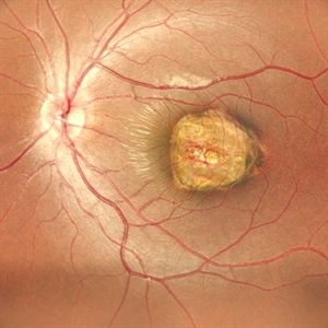
Female 19-years-old with tuberous sclerosis. BCVA LE 10/10. Fundus LE shows three multinodular hamartomas.
Photographer: Sofia Sousa Mano
Imaging device: Canon CR 2 plus
Condition/keywords: hamartoma, tuberculous chorioretinitis
-
 Intraocular Foreign Body
Feb 7 2019 by Somnath Chakraborty, MD
Left eye fundus photo montage of a 45-year-old male showing a large iron foreign body, impacted inferior to the infero-temporal branch vessels with a large patch of surrounding chorio-retinal atrophy, secondary to resolving Commotio retinae
Photographer: Saptarshi Mehta
Condition/keywords: commotio retinae, intraocular foreign body, trauma
-
 ICG: Choroidal Aspergilloma With Secondary Choroidal Neovascularization and Exudative Retinal Detachment
Mar 21 2019 by Scott D Walter, MD, MSc, FASRS
Multimodal imaging of a transplant patient with disseminated Aspergillosis and vision loss in her left eye.
Condition/keywords: choroidal neovascular membrane (CNVM), choroidal neovascularization (CNV), exudative detachment, focal chorioretinitis, fungal endophthalmitis, granulomatous choroiditis
-
 Retinochoroidal Coloboma With Aberrant Vasculature
Nov 10 2018 by Chintan D Desai, MBBS, DO, DNB, FICO
Fundus photo montage of a 32-year-old female with a retinochoroidal coloboma Ida Mann classification type 3 with a spring coil shaped aberrant vessel.
Photographer: Kankan Talukdar
Imaging device: Zeiss FF4
Condition/keywords: chorioretinal coloboma
-
 Chorio-Retinal Coloboma
Aug 4 2017 by Marco D'Angelo
Left eye, 7-year-old female patient, normal visual acuity,
Photographer: Dr. Marco D'Angelo, S.Chiara Hospital, Trento, Italy
Imaging device: Topcon TRC-NW 6S
Condition/keywords: coloboma
-
 Multifocal CSR FA & ICG
May 19 2017 by Manish Nagpal, MD, FRCS (UK), FASRS
A 30-year-old male diagnosed elsewhere as VKH was started on heavy steroids and he developed multiple serous elevations and OS developed a exudative RD. We immediately asked the patient to stop steroids and when he followed up after a month lesions had flattened and he had recovered to 20/40 in both eyes.. he is still undergoing further follow up at this stage...
Photographer: pooja barot
Imaging device: heidelberg
Condition/keywords: central serous retinopathy (CSR), multifocal central serous chorioretinopathy (CSCR), Vogt-Koyanagi-Harada
-
 Central Serous Chorioretinopathy
Apr 19 2017 by Gustavo Barreto de Melo, MD, PhD, FASRS
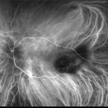
Fundus photograph of a 32-year-old pregnant woman with a serous detachment and subretinal fibrin deposit surrounding the fovea.
Photographer: Denyson Silva, Sergipe Eye Hospital
Condition/keywords: central serous chorioretinopathy (CSCR), pregnancy
-
 Toxoplasmosis Associated Epiretinal Membrane
Oct 27 2016 by Gabriel Costa Andrade, PhD
Fundus photograph of a 26-year-old woman with a chorioretinal scar due to toxoplasmosis and secondary epiretinal membrane.
Photographer: Gabriel Andrade, Federal University of São Paulo, São Paulo, Brazil
Condition/keywords: epiretinal membrane (ERM), toxoplasmosis
 Loading…