Search results (959 results)
-
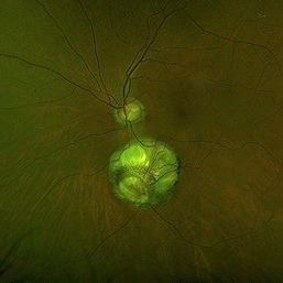
 Franceschetti's Sign
Jun 5 2025 by César Adrián Gómez Valdivia, MD
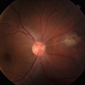
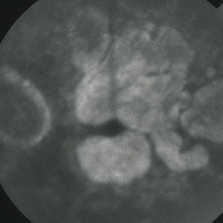
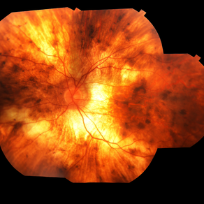
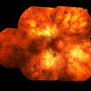
Franceschetti's sign found in a 22 year-old female patient diagnosed with ocular toxoplasmosis. These bands typically link an old scar to the optic disc, indicative of previous inflammation. Findings were unilateral.
Photographer: @eyemissu2
Imaging device: TOPCON TRC-50DX
Condition/keywords: chorio, Franceschetti's Sign, toxoplasmosis
-
 "Starry Sky" Fundus in Vogt-Koyanaki-Harada Syndrome
Jan 10 2018 by Peter H. Tang, MD, PhD
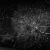
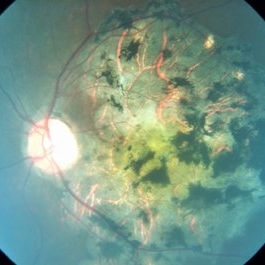
Fluorescein angiography imaging of a 27-year-old male with acute inflammation as part of Vogt-Koyanagi-Harada Syndrome.
Imaging device: Optos California
Condition/keywords: chorioretinal inflammations, retina, uveitis, Vogt-Koyanagi-Harada
-
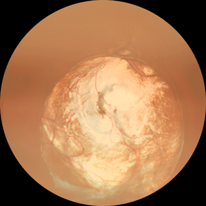
 Acute syphilitic posterior placoid chorioretinitis
Apr 24 2022 by Aniruddha K Agarwal, MD
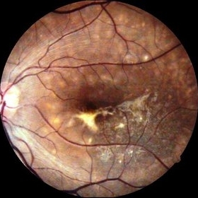
Green-light fundus autofluorescence (FAF) of the right eye from a 55-year-old man with risk factors for sexually trasnmitted diseases who presented to the retina clinic for a central scotoma. Funduscopy revealed a placoid lesion in the posterior pole. FAF highlights a hyperautofluorescent placoid lesion involving the macula with granular hyperfluorescence. The patient tested positive for syphilis and received intravenous penicillin treatment.
Photographer: Esther CIANCAS, MD, PhD, Gema CRESPO-RODRÍGUEZ, RN
Imaging device: Zeiss Clarus fundus camera
Condition/keywords: chorioretinitis, IUSG, syphilis, uveitis
-
 Anastomosis
Jul 29 2025 by Drew Mitchell
3x3 OCT-Angiography Full Depth Color Coded of a left eye with Macular Telangiectasia Type 2
Photographer: Drew Mitchell, OCT-C
Imaging device: Zeiss Cirrus 5000
Condition/keywords: chorioretinal anastomosis, macular telangiectasia type 2, retinochoroidal anastomosis
-
 BB Gun Accident
Dec 13 2017 by Jason Griffith
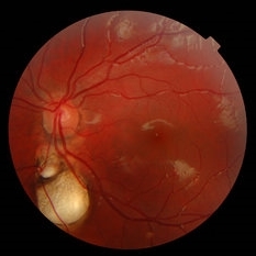
8-year-old male patient presented with history of metallic extraocular foreign body OD x 3 weeks; patient voiced history of being struck by BB approximately 3 weeks prior to exam; pt. was taken to OR for removal of metallic intraocular foreign body.
Photographer: Jason Griffith
Imaging device: Optos California
Condition/keywords: chorioretinitis sclopetaria
-
---thumb.jpg/image-square;max$300,300.ImageHandler) case 2 OD
case 2 OD
Feb 14 2013 by From the Collections of Thomas M. Aaberg, MD and Thomas M. Aaberg Jr., MD
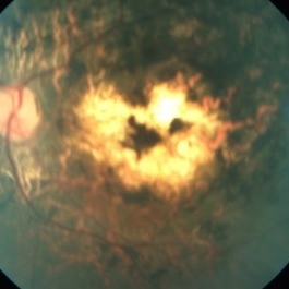
reproductions of figures 4 and 5 from the article "Ocular involvement in neonatal herpes simplex virus infection" (Hagler WS et al, Arch Opthalmol 1969;82:169-76.). Fulminating chorioretinal scarring and retinal pigmentary changes were seen in both eyes of an infant with neonatal systemic herpesvirus infection.
Condition/keywords: chorioretinal scar, neonatal herpes
-
 Central Areolar Choriocapillaris Atrophy
Mar 26 2019 by Gary R. Cook, MD, FACS
Right eye of a 64-year-old male with central (or regional) atrophy of the RPE and choriocapillaris in the macula; VA= 20/30.
Imaging device: Topcon VT-50
Condition/keywords: choriocapillaris, hereditary choroidal atrophy, hereditary choroidal dystrophy
-
 Central Areolar Choriocapillaris Atrophy
Mar 26 2019 by Gary R. Cook, MD, FACS
Left eye of a 64-year-old male with central (regional) areolar choroidal dystrophy showing fairly well circumscribed atrophy of the RPE and choriocapillaris in the macula; VA= 20/30
Imaging device: Topcon VT-50
Condition/keywords: choriocapillaris, hereditary choroidal atrophy, hereditary choroidal dystrophy
-
 Central Areolar Choriocapillaris Atrophy
Mar 26 2019 by Gary R. Cook, MD, FACS
Late-phase fluorescein angiogram image of the left eye of a 64-year-old white male with central areolar choriocapillaris atrophy showing light late staining of the central lesions OS; V.A. = 20/30
Imaging device: Topcon VT-50
Condition/keywords: choriocapillaris, FA late phase, fluorescein angiogram (FA), hereditary choroidal atrophy, hereditary choroidal dystrophy
-
 Chorio Retinal Atrophy
Apr 23 2015 by Mehul A Shah
Patient presented with progressive visual loss ou.
Photographer: Mehul Shah
Condition/keywords: chorioretinal atrophy
-
 Chorio Retinal Scar
Feb 19 2024 by Sanauddin Samejo , Diploma (Ophthalmic Technician Training Course)
A patient came in to Clinic of Dr Madhav Rao (VR Surgeon)
Photographer: Sanauddin Samejo, Burjeel Hospital, Abu Dhabi, UAE.
Imaging device: Optos Silverstone
Condition/keywords: chorioretinal scar
-
 Chorio Retinital Inflammation
Sep 21 2014 by Mehul A Shah
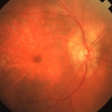
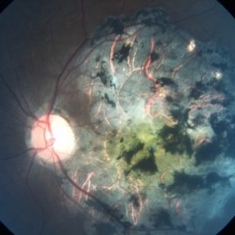
A 45-year-old female presented with complaints of gradual loss of vision before 3 months and when reached to hospital had this picture.
Photographer: Drashti Netralaya,Dahod
Imaging device: Zeiss ff450
Condition/keywords: chorioretinal inflammations
-
 Chorio-Retinal Coloboma
Aug 4 2017 by Marco D'Angelo
Left eye, 7-year-old female patient, normal visual acuity,
Photographer: Dr. Marco D'Angelo, S.Chiara Hospital, Trento, Italy
Imaging device: Topcon TRC-NW 6S
Condition/keywords: coloboma
-
 Choriodal Rupture 004 - Fundus Autoflurescence - 6 Month Follow Up
Mar 11 2013 by Suber S. Huang, MD, MBA, FASRS
40-year-old male sustained blunt trauma OD with orbital fracture and choroidal rupture subjacent to inferior arcade with blood, subretinal fluid, and exudate extending to fovea with CF 2 feet at presentation 9-10-12. On follow up 3-11-12, vision improved to 20/400 with resolutionof hemorrhage, normal OCT, but speckling of foveal RPE and PMB consistent with damage.
Photographer: Mark Harrod
Condition/keywords: autofluorescence imaging, blunt trauma, choroidal hemorrhage, choroidal rupture, orbital fracture, retinal hemorrhage, submacular hemorrhage
-
 Choriodal Rupture - 003 - 6 Month Follow Up
Mar 11 2013 by Suber S. Huang, MD, MBA, FASRS
40 -year-old male sustained blunt trauma OD with orbital fracture and choroidal rupture subjacent to inferior arcade with blood, subretinal fluid, and exudate extending to fovea with CF 2 feet at presentation 9-10-12. On followup 3-11-12, vision improved to 20/400 with resolutionof hemorrhage, normal OCT, but speckling of foveal RPE and PMB consistent with damage.
Photographer: Mark Harrod
Condition/keywords: autofluorescence imaging, blunt trauma, choroidal hemorrhage, choroidal rupture, orbital fracture, retinal hemorrhage, submacular hemorrhage
-
 Choriodal Rupture - AW 001 - Initial Presentation
Mar 11 2013 by Suber S. Huang, MD, MBA, FASRS
40-year-old male sustained blunt trauma OD with orbital fracture and choroidal rupture subjacent to inferior arcade with blood, subretinal fluid, and exudate extending to fovea with CF 2 feet at presentation 9-10-12. On followup 3-11-12, vision improved to 20/400 with resolution of hemorrhage, normal OCT, but speckling of foveal RPE and PMB consistent with damage.
Photographer: Mark Harrod
Condition/keywords: autofluorescence imaging, blunt trauma, choroidal hemorrhage, choroidal rupture, orbital fracture, retinal hemorrhage, submacular hemorrhage
-
 Choriodal Rupture - AW 002 - 6 Month F/U
Mar 11 2013 by Suber S. Huang, MD, MBA, FASRS
40-year-old male sustained blunt trauma OD with orbital fracture and choroidal rupture subjacent to inferior arcade with blood, subretinal fluid, and exudate extending to fovea with CF 2 feet at presentation 9-10-12. On follow up 3-11-12, vision improved to 20/400 with resolutionof hemorrhage, normal OCT, but speckling of foveal RPE and PMB consistent with damage.
Photographer: Mark Harrod
Condition/keywords: autofluorescence imaging, blunt trauma, choroidal hemorrhage, choroidal rupture, orbital fracture, retinal hemorrhage, submacular hemorrhage
-
 Choriodemia
Jul 8 2013 by David W. Faber, MD
Fundus montage of a 50-year-old male.
Photographer: Donna Knight C.R.A., Rocky Mountain Retina Consultants, Sale Lake City, Utah
Condition/keywords: choroideremia
-
 Chorioderemia
Jul 8 2013 by David W. Faber, MD
Fundus montage of a 50-year-old male.
Photographer: Donna Knight, C.R.A, Rocky Mountain Retina Consultants, Salt Lake City, Utah
Condition/keywords: choroideremia
-
 Chorioretinal Atrophy
Oct 1 2014 by Mehul A Shah
A 25-year-old male patient presented with bilateral similar picture.
Photographer: Drashti Netralaya,Dahod
Imaging device: Zeiss ff450
Condition/keywords: chorioretinal atrophy
-
 Chorioretinal Atrophy
Oct 2 2017 by Mehul A Shah
A 24-year-old female presented to is with complaint of gradual loss of vision.
Photographer: Mehul Shah
Condition/keywords: chorioretinal atrophy
-
 Chorioretinal Coloboma
Aug 7 2023 by Aditya S Kelkar, MS, FRCS, FASRS,FRCOphth
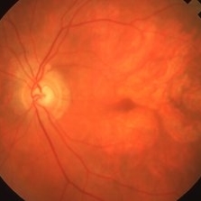
Fundus photograph of an 68-year-old woman with a chorioretinal coloboma observed.
Photographer: Optom Komal Jangam, National Institute of Ophthalmology, Pune, India.
Imaging device: OPTOS DAYTONA
Condition/keywords: chorioretinal coloboma
-
 Chorioretinal Coloboma
May 2 2023 by RAKESH SHAH, MS DNB FACS FRF FICO MBA
Young man with blurring of vision in both eyes
Photographer: Dr.Rakesh shah
Condition/keywords: chorioretinal coloboma
-
 Chorioretinal coloboma 1
Jan 11 2013 by Alex P. Hunyor, MD
Inferior chorioretinal coloboma - color image 1.
Condition/keywords: chorioretinal coloboma, coloboma of choroid
-
 Chorioretinal coloboma 2
Jan 11 2013 by Alex P. Hunyor, MD
Inferior chorioretinal coloboma - color image 2.
Condition/keywords: chorioretinal coloboma, coloboma of choroid
 Loading…