Search results (64 results)
-
 Vascular Non Perfusion in Takayasu Arteritis
Feb 6 2024 by SHILPI H NARNAWARE, ICO ( Retina) , FAICO ( Vitreo-Retina)
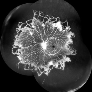
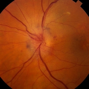
A case of 16 year-old female with combined RD in RE. Fundus examination & FFA revealed 360 degrees non-perfusion in periphery in non-symptomatic eye.
Photographer: Shilpi Narnaware, Sarakshi Netralaya , Nagpur, Maharashtra , India
Imaging device: Mirante ( by Nidek)
Condition/keywords: CNP areas, takayasu arteritis
-
 Takayasu Retinopathy
Apr 30 2025 by Vishal Agrawal, MD, FRCS,FACS,FASRS
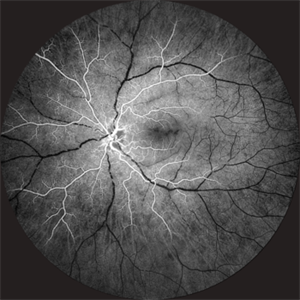
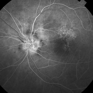
Fundus fluorescein angiography image of a young girl with diagnosed Takayasu arteritis who presented with complains of diminished vision in both eyes. FFA shows complete absence of venous filling with segmented blood column secondary to CRAO with peripheral avascular area.
Photographer: Dr Ayushi Gupta
Imaging device: Clarus 700
Condition/keywords: calcified drusen, CRAO, takayasu arteritis
-
 Susac's Syndrome
Feb 13 2018 by John S. King, MD
Background: 46-year-old WF with CML (stable on Sprycel) saw her PCP for headaches without known cause; Headaches worsened and became confused, disoriented, off balance, and impaired short term memory. Heme-oncology ordered MRI that showed abnormal signal in the cerebellum and other parts of the brain, and LP has elevated protein. LP did show positive tau test, but fortunately, was a false positive for CJD. IV and PO steroids started and symptoms improved. MRI showed much improvement one month since starting steroids. 3 weeks later had a scotoma in right eye and eye doctor did not find anything at that time to cause it. Tinnitus developed (and some intermittent vertigo before that) and ENT referred back to eye doctor, who then referred the patient to Dr. Zocchi. He found a CWS and BRAO OD, and bilateral arteritis. She had some additional work-up for vasculitis. Given the retinal arteritis, cochlear issues, and MRI findings, Dr.Zocchi suspected Susac's Syndrome. She was started on multiple regimens including prednisone, IVIG, azathiprine, and MTX, and has had the best reponse to IVIG (FA shows a recurrence/worsening while adjusting IMT). She is stable and doing well with 20/20 vision in both eyes.
Photographer: Kay Dalby
Imaging device: Topcon
Condition/keywords: retinal vasculitis, Susac's syndrome
-
 Takayasu Retinopathy
Apr 30 2025 by Vishal Agrawal, MD, FRCS,FACS,FASRS
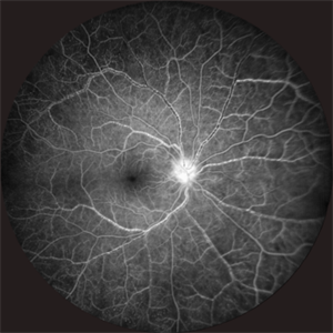
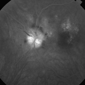
Fundus fluorescein angiography image of a young girl with diagnosed Takayasu arteritis who presented with complains of diminished vision in both eyes. FFA shows complete absence of venous filling with segmented blood column secondary to CRAO with peripheral avascular area.
Photographer: Dr Ayushi Gupta
Imaging device: Clarus 700
Condition/keywords: CRAO, Takayasus disease
-
 Anterior Ischemic Optic Neuropathy and Choroidal Ischemia
Mar 1 2014 by Homayoun Tabandeh, MD, FASRS
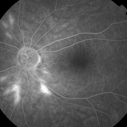
Arteritic anterior ischemic optic neuropathy and choroidal ischemia in a patient with giant cell arteritis.
Condition/keywords: anterior ischemic optic neuropathy, giant cell arteritis
-
 Anterior Ischemic Optic Neuropathy and Choroidal Ischemia
Mar 1 2014 by Homayoun Tabandeh, MD, FASRS
Fundus fluorescein angiogram of a patient with arteritic anterior ischemic optic neuropathy and choroidal ischemia associated with giant cell arteritis.
Condition/keywords: anterior ischemic optic neuropathy
-
 Anterior ischemic optic neuropathy slide 1
Oct 22 2012 by Ronald C. Gentile, MD
70-year-old women with acute loss of vision in the left eye. Review of symptoms was significant for temporal arteritis and ESR was very high. Fundus examination of the left eye had a swollen white optic nerve head with a few peri-papillary cotton wool spots.
Photographer: The New York Eye & Ear Infirmary Department of Medical Imaging
Condition/keywords: anterior ischemic optic neuropathy, choroidal ischemia, temporal arteritis
-
 Anterior ischemic optic neuropathy slide 2
Oct 22 2012 by Ronald C. Gentile, MD
Early fluorescein angiography revealed delayed filling of the choroid and optic nerve.
Photographer: The New York Eye & Ear Infirmary Department of Medical Imaging
Condition/keywords: anterior ischemic optic neuropathy, choroidal ischemia, temporal arteritis
-
 Anterior ischemic optic neuropathy slide 3
Oct 22 2012 by Ronald C. Gentile, MD
Late fluorescein angiography revealed leakage of the optic disc.
Photographer: The New York Eye & Ear Infirmary Department of Medical Imaging
Condition/keywords: anterior ischemic optic neuropathy, temporal arteritis
-
 Arteritis
Apr 18 2013 by Howard Schatz, MD
III arteritis, right eye: 20/20; left eye: 4/200.
Condition/keywords: arteritis
-
 Biopsy Proven Giant Cell Arteritis
Oct 15 2018 by Darin R. Goldman, MD
83-year-old male with biopsy-proven giant cell arteritis OU and old BRVO OS.
Photographer: Crystal Esparza, BS, COA, Retina Group of Florida
Imaging device: Topcon TRC 50DX
Condition/keywords: branch retinal vein occlusion (BRVO), giant cell arteritis, optic disc edema, papilledema
-
 BRAMAD
Apr 23 2013 by Howard Schatz, MD
III BRAMAD.
Condition/keywords: bilateral retinal arteritis with multiple aneurysmal dilatation (BRAMAD)
-
 BRAMAD
Apr 23 2013 by Howard Schatz, MD
III BRAMAD.
Condition/keywords: bilateral retinal arteritis with multiple aneurysmal dilatation (BRAMAD)
-
 BRAMAD
Apr 23 2013 by Howard Schatz, MD
III BRAMAD.
Condition/keywords: bilateral retinal arteritis with multiple aneurysmal dilatation (BRAMAD)
-
 BRAMAD
Apr 23 2013 by Howard Schatz, MD
41-year-old female, III BRAMAD, 20/400; 20/15.
Condition/keywords: bilateral retinal arteritis with multiple aneurysmal dilatation (BRAMAD)
-
 BRAMAD
Apr 23 2013 by Howard Schatz, MD
III BRAMAD, 35-year-old female, right eye: 20/60; left eye: 20/20.
Condition/keywords: bilateral retinal arteritis with multiple aneurysmal dilatation (BRAMAD)
-
 BRAMAD
Apr 23 2013 by Howard Schatz, MD
III BRAMAD.
Condition/keywords: bilateral retinal arteritis with multiple aneurysmal dilatation (BRAMAD)
-
 BRAMAD
Apr 23 2013 by Howard Schatz, MD
42-year-old female, III BRAMAD.
Condition/keywords: bilateral retinal arteritis with multiple aneurysmal dilatation (BRAMAD)
-
 BRAMAD
Apr 23 2013 by Howard Schatz, MD
III BRAMAD.
Condition/keywords: bilateral retinal arteritis with multiple aneurysmal dilatation (BRAMAD)
-
 BRAMAD
Apr 23 2013 by Howard Schatz, MD
38-year-old white female, II BRAMAD.
Condition/keywords: bilateral retinal arteritis with multiple aneurysmal dilatation (BRAMAD)
-
 BRAMAD
Apr 23 2013 by Howard Schatz, MD
38-year-old white female, III BRAMAD, right eye: 20/25, HM.
Condition/keywords: bilateral retinal arteritis with multiple aneurysmal dilatation (BRAMAD)
-
 BRAMAD
Apr 23 2013 by Howard Schatz, MD
23-year-old white female, III BRAMAD.
Condition/keywords: bilateral retinal arteritis with multiple aneurysmal dilatation (BRAMAD)
-
---thumb.jpg/image-square;max$300,300.ImageHandler) Brown/Mendis BJO 57:344, 1973
Brown/Mendis BJO 57:344, 1973
Feb 14 2013 by From the Collections of Thomas M. Aaberg, MD and Thomas M. Aaberg Jr., MD
reprints of figures 1 and 2 from the publication Brown and Mendis. Retinal arteritis complicating herpes zoster ophthalmicus. Br J Ophthalmol 1973;57:344-6. The left panel is a "fundus painting showing extensive exudate in areas of supply of narrowed and sheathed upper nasal and upper temporal retinal arterioles." The right panel is a fluorescein angiograph of the fundus, "demonstrating leakage of dye in area of exudation."
Condition/keywords: Herpes zoster, retinal arteriolar occlusion, retinal necrosis
-
 Combined GCA and AH
Oct 27 2020 by Nathan C. Steinle, MD
This patient demonstrates two rare diseases in one image: giant cell arteritis choroidal vascular ischemia with asteroid hyalosis. Delayed choroidal filling can be a presenting sign of giant cell arteritis, which is a life-threatening disease.
Photographer: Nancy Gutierrez
Imaging device: Optos
Condition/keywords: asteroid hyalosis, giant cell arteritis
 Loading…