Search results (64 results)
-
 Arteritis
Apr 18 2013 by Howard Schatz, MD
III arteritis, right eye: 20/20; left eye: 4/200.
Condition/keywords: arteritis
-
 Slide 1-27
Feb 19 2019 by Lancaster Course in Ophthalmology
Acute arteritis in an orbital inflammatory pseudotumor. Arterial wall is edematous and thickened with PMNs, lymphocytes, and macrophages. Eosinophils are seen in the tissue at right. (H&E stain)
Condition/keywords: arteritis, edematous, eosinophils, polymorphonuclear leukocytes (PMNs), pseudotumor
-
 Anterior Ischemic Optic Neuropathy and Choroidal Ischemia
Mar 1 2014 by Homayoun Tabandeh, MD, FASRS
Arteritic anterior ischemic optic neuropathy and choroidal ischemia in a patient with giant cell arteritis.
Condition/keywords: anterior ischemic optic neuropathy, giant cell arteritis
-
 Anterior Ischemic Optic Neuropathy and Choroidal Ischemia
Mar 1 2014 by Homayoun Tabandeh, MD, FASRS
Fundus fluorescein angiogram of a patient with arteritic anterior ischemic optic neuropathy and choroidal ischemia associated with giant cell arteritis.
Condition/keywords: anterior ischemic optic neuropathy
-
 Anterior ischemic optic neuropathy slide 1
Oct 22 2012 by Ronald C. Gentile, MD
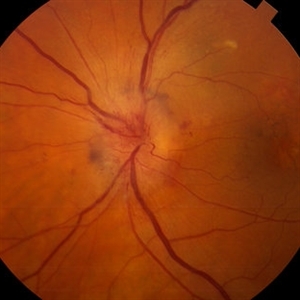
70-year-old women with acute loss of vision in the left eye. Review of symptoms was significant for temporal arteritis and ESR was very high. Fundus examination of the left eye had a swollen white optic nerve head with a few peri-papillary cotton wool spots.
Photographer: The New York Eye & Ear Infirmary Department of Medical Imaging
Condition/keywords: anterior ischemic optic neuropathy, choroidal ischemia, temporal arteritis
-
 Anterior ischemic optic neuropathy slide 2
Oct 22 2012 by Ronald C. Gentile, MD
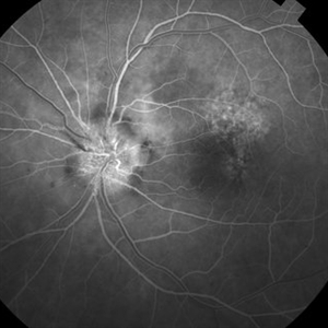
Early fluorescein angiography revealed delayed filling of the choroid and optic nerve.
Photographer: The New York Eye & Ear Infirmary Department of Medical Imaging
Condition/keywords: anterior ischemic optic neuropathy, choroidal ischemia, temporal arteritis
-
 Anterior ischemic optic neuropathy slide 3
Oct 22 2012 by Ronald C. Gentile, MD
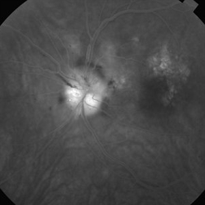
Late fluorescein angiography revealed leakage of the optic disc.
Photographer: The New York Eye & Ear Infirmary Department of Medical Imaging
Condition/keywords: anterior ischemic optic neuropathy, temporal arteritis
-
 Biopsy Proven Giant Cell Arteritis
Oct 15 2018 by Darin R. Goldman, MD
83-year-old male with biopsy-proven giant cell arteritis OU and old BRVO OS.
Photographer: Crystal Esparza, BS, COA, Retina Group of Florida
Imaging device: Topcon TRC 50DX
Condition/keywords: branch retinal vein occlusion (BRVO), giant cell arteritis, optic disc edema, papilledema
-
 BRAMAD
Apr 23 2013 by Howard Schatz, MD
III BRAMAD.
Condition/keywords: bilateral retinal arteritis with multiple aneurysmal dilatation (BRAMAD)
-
 BRAMAD
Apr 23 2013 by Howard Schatz, MD
III BRAMAD.
Condition/keywords: bilateral retinal arteritis with multiple aneurysmal dilatation (BRAMAD)
-
 BRAMAD
Apr 23 2013 by Howard Schatz, MD
III BRAMAD.
Condition/keywords: bilateral retinal arteritis with multiple aneurysmal dilatation (BRAMAD)
-
 BRAMAD
Apr 23 2013 by Howard Schatz, MD
41-year-old female, III BRAMAD, 20/400; 20/15.
Condition/keywords: bilateral retinal arteritis with multiple aneurysmal dilatation (BRAMAD)
-
 BRAMAD
Apr 23 2013 by Howard Schatz, MD
III BRAMAD, 35-year-old female, right eye: 20/60; left eye: 20/20.
Condition/keywords: bilateral retinal arteritis with multiple aneurysmal dilatation (BRAMAD)
-
 BRAMAD
Apr 23 2013 by Howard Schatz, MD
III BRAMAD.
Condition/keywords: bilateral retinal arteritis with multiple aneurysmal dilatation (BRAMAD)
-
 BRAMAD
Apr 23 2013 by Howard Schatz, MD
42-year-old female, III BRAMAD.
Condition/keywords: bilateral retinal arteritis with multiple aneurysmal dilatation (BRAMAD)
-
 BRAMAD
Apr 23 2013 by Howard Schatz, MD
III BRAMAD.
Condition/keywords: bilateral retinal arteritis with multiple aneurysmal dilatation (BRAMAD)
-
 BRAMAD
Apr 23 2013 by Howard Schatz, MD
38-year-old white female, II BRAMAD.
Condition/keywords: bilateral retinal arteritis with multiple aneurysmal dilatation (BRAMAD)
-
 BRAMAD
Apr 23 2013 by Howard Schatz, MD
38-year-old white female, III BRAMAD, right eye: 20/25, HM.
Condition/keywords: bilateral retinal arteritis with multiple aneurysmal dilatation (BRAMAD)
-
 BRAMAD
Apr 23 2013 by Howard Schatz, MD
23-year-old white female, III BRAMAD.
Condition/keywords: bilateral retinal arteritis with multiple aneurysmal dilatation (BRAMAD)
-
---thumb.jpg/image-square;max$300,300.ImageHandler) Brown/Mendis BJO 57:344, 1973
Brown/Mendis BJO 57:344, 1973
Feb 14 2013 by From the Collections of Thomas M. Aaberg, MD and Thomas M. Aaberg Jr., MD
reprints of figures 1 and 2 from the publication Brown and Mendis. Retinal arteritis complicating herpes zoster ophthalmicus. Br J Ophthalmol 1973;57:344-6. The left panel is a "fundus painting showing extensive exudate in areas of supply of narrowed and sheathed upper nasal and upper temporal retinal arterioles." The right panel is a fluorescein angiograph of the fundus, "demonstrating leakage of dye in area of exudation."
Condition/keywords: Herpes zoster, retinal arteriolar occlusion, retinal necrosis
-
 Combined GCA and AH
Oct 27 2020 by Nathan C. Steinle, MD
This patient demonstrates two rare diseases in one image: giant cell arteritis choroidal vascular ischemia with asteroid hyalosis. Delayed choroidal filling can be a presenting sign of giant cell arteritis, which is a life-threatening disease.
Photographer: Nancy Gutierrez
Imaging device: Optos
Condition/keywords: asteroid hyalosis, giant cell arteritis
-
---thumb.jpg/image-square;max$300,300.ImageHandler) Foci of arteriolar plaques
Foci of arteriolar plaques
Feb 15 2013 by From the Collections of Thomas M. Aaberg, MD and Thomas M. Aaberg Jr., MD
color fundus photograph showing foci of arteriolar plaques (so-called Kyrieleis arteritis), as seen in ocular toxoplasmosis.
Condition/keywords: ocular toxoplasmosis
-
---thumb.jpg/image-square;max$300,300.ImageHandler) Foci of arteriolar plaques proximal to an area of retinal whitening consistent with ocular toxoplasmosis
Foci of arteriolar plaques proximal to an area of retinal whitening consistent with ocular toxoplasmosis
Feb 15 2013 by From the Collections of Thomas M. Aaberg, MD and Thomas M. Aaberg Jr., MD
Color fundus photograph showing foci of arteriolar plaques (so-called Kyrieleis arteritis) proximal to an area of retinal whitening consistent with ocular toxoplasmosis.
Condition/keywords: ocular toxoplasmosis
 Loading…