Search results (87 results)
-
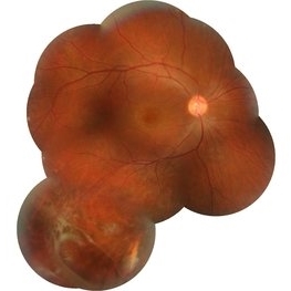
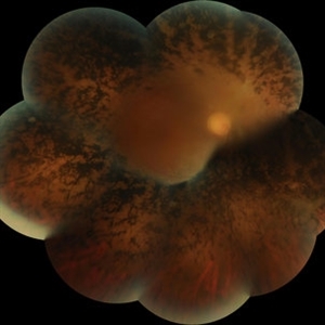
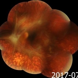 Advanced Proliferative Diabetic Retinopathy
Nov 4 2017 by Hamid Ahmadieh, MD
Merged color fundus photograph of the left eye of a 30-year-old woman with type1 diabetes since childhood. Note laser scars, severe fibrous proliferation, traction RD and macular dragging.
Photographer: Shabnam Poureh, Negah Eye Center, Tehran, Iran
Condition/keywords: color fundus photograph, diabetes, fibrous proliferation, proliferative diabetic retinopathy (PDR), severe traction
-
 Branch Retinal Vein Occlusion- Fluorescein Angiogram, Montage
Apr 15 2016 by James B. Soque, CRA, OCT-C, COA, FOPS
A fluorescein angiogram of an 80-year-old white female with a superotemporal branch retinal vein occlusion, and retinal edema of the right eye. Currently receiving Lucentis 0.5 injection therapy.
Photographer: James Soque, CRA OCT-C COA, Island Retina, Shirley, NY
Imaging device: Topcon TRC, MERGE Imaging Software V. 11.2.0
Condition/keywords: branch retinal vein occlusion (BRVO), montage, non-perfused branch retinal vein occlusion (BRVO)
-
 Central retinal artery occlusion
Nov 30 2022 by Ethan K Sobol, MD
A central retinal artery occlusion with cilioretinal artery sparing, imaged using a Volk Panretinal 2.2 and an iPhone camera in the emergency department.
Photographer: Jared Raabe, MD, Emory University Hospital
Imaging device: IPhone 13 Pro
Condition/keywords: central retinal artery occlusion (CRAO)
-
 CSCR Mushroom Cloud
Feb 23 2015 by James J. Bedrick, MD
Late transit FA of a large active sub-foveal CSCR leak. You may view this pair in stereo to appreciate the plume of leakage within this large serous RD of the macula. This patient presented with a BCVA of 20/200 and fluorescein and historic evidence of prior episodes of leakage. After discussion of known treatment options including observation, he elected to be treated initially with oral rifampin and BCVA improved to 20/40 with persistent metamorphosis and a shallower persistent macular detachment over several visits. Rifampin was discontinued and he then received sub-threshold micro-pulse laser photocoagulation with an 810 diode which resulted in the patient reporting full restoration of his vision subjectively within a month. He failed to keep his follow-up appointment.
Photographer: Diana Bodnar, COT
Imaging device: Topcon 50X with Merge capture station
Condition/keywords: CSCR subfoveal leak
-
 Diabetic Macular Edema, Proliferative Diabetic Retinopathy, Neovascularization Elsewhere, DME, PDR, NVE
Apr 1 2013 by James B. Soque, CRA, OCT-C, COA, FOPS
39-year-old white female and long standing diabetis, c/o new peripheral symptoms of left eye. FA OS reveals diabetic macular edema, microaneurysms, and neovasculaization elsewhere. Fluorescein Angogram, Early Phase, 50 Deg, 2x Mag.
Photographer: James B Soque, CRA, COA
Imaging device: Topcon TRC 50DX with MERGE software, OIS 10.6.45
Condition/keywords: diabetic macular edema, neovascularization (NV), proliferative diabetic retinopathy (PDR)
-
 Diabetic Retinopathy, CSME, Exudates, NVD, Color Fundus Photo, Montage
Mar 18 2015 by James B. Soque, CRA, OCT-C, COA, FOPS
A 58-year-old diabetic male with a longstanding history of diabetic eye disease. Left eye color fundus photo shows extensive CSME, Clinically Significant Macular Edema, with deposits of hard exudates at fixation. There is extensive scattering of hard exudates and sheathing of the vessels.
Photographer: James B Soque, CRA COA
Imaging device: Topcon TRC 50 DX, OIS 5 MP Camera, MERGE software
Condition/keywords: background diabetic retinopathy (BDR), creamy yellow exudates, diabetes, exudates over the posterior pole, neovascularization of the disc (NVD), vessel sheathing
-
 Fibrovascular Retinal Pigment Epithelial Detachment - Color Fundus
Jul 16 2014 by James B. Soque, CRA, OCT-C, COA, FOPS
69-year-old white female with Hx of 10 anti-VEFG treatment injections of right eye, VA 20/200, now stable, off drug for 10 months.
Photographer: James B Soque, CRA COA
Imaging device: Topcon TRC 50 DX with MERGE software, 5 MP dig camera
Condition/keywords: color fundus photograph, fibrovascular pigment epithelial detachment (PED), pigment epithelial atrophy, retina
-
 Macular Star
Sep 8 2024 by Cesar Augusto Rocha Rojas, MD
Fundus photograph of a 27-year-old male with hypertensive emergency secondary to chronic kidney disease.
Photographer: Cesar Augusto Rocha Rojas, Hospital General de Zona #20, Instituto Mexicano del Seguro Social (IMSS)
Imaging device: Smartphone, Pan Retinal 2.2 Lens
Condition/keywords: macular star
-
 Subconjuntival IOL After Blunt Trauma
Jun 27 2018 by Gabriel Costa Andrade, PhD
A 73-year-old male patient was referred to our ophthalmic emergency department with complaints of redness, pain, and diminution of vision in his left eye, after fall from height. The patient underwent small incision cataract surgery with polymethylmethacrylate (PMMA) IOL implantation in both the eyes 15 years back through superior sclerocorneal incision under local anesthesia. His best-corrected visual acuity was perception of light in the left eye. Ophthalmic examination using slit lamp biomicroscopy of the left eye revealed diffuse subconjunctival hemorrhage with no conjunctival laceration and inferior bulbar conjunctiva showed traumatic pseudophacocele with a sign “golden half ring,” suggesting the presence of PCIOL in subconjunctival space.There was total hyphema obscuring the view of rest of the ocular structures in his left eye.
Photographer: Gabriel Andrade, RETINA CLINIC, São Paulo, BRAZIL
Condition/keywords: dislocated intraocular lens (IOL), trauma
-
 Extra-scleral Extension of Choroidal Melanoma
Dec 23 2021 by Jessica Norkus
89-year-old female with extra-scleral extension of choroidal metastatic melanoma. Patient hadn't been seen by any eye doctor in 3 years prior to this visit. Noticed scleral darkening about 6 months ago, with vision loss noted for about 4-5 months. Presented with LP vision. Emergent MRI of brain/orbit showed no extension beyond what is seen at slit lamp. CT C/A/P w/ contrast ordered and found 2 hepatic lesions, concerning for potential mets. Patient referred to medical oncology.
Photographer: Jessica Norkus, COA, OSC
Imaging device: Topcon TRC 50DX
Condition/keywords: external photography, extrascleral extension, metastatic cancer, metastatic lesion
-
 Choroidal Melanoma
Jan 8 2016 by Jared Watson
42-year-old white female, S/P I-125 plaque brachytherapy.
Photographer: Jared Watson COT/CRA
Imaging device: Topcon 50EX, OIS-Merge
-
 Extra-scleral Extension of Choroidal Melanoma
Dec 23 2021 by Jessica Norkus
89-year-old female with extra-scleral extension of choroidal metastatic melanoma. Patient hadn't been seen by any eye doctor in 3 years prior to this visit. Noticed scleral darkening about 6 months ago, with vision loss noted for about 4-5 months. Presented with LP vision. Emergent MRI of brain/orbit showed no extension beyond what is seen at slit lamp. CT C/A/P w/ contrast ordered and found 2 hepatic lesions, concerning for potential mets. Patient referred to medical oncology.
Photographer: Jessica Norkus, COA, OSC
Imaging device: Topcon TRC 50DX
Condition/keywords: extrascleral extension, metastatic cancer, metastatic lesion
-
 Juvenile Retinoschisis
Oct 10 2015 by Hamid Ahmadieh, MD
Merged color fundus photograph of the right eye of a 30-year-old man with juvenile retioschisis. Involvement of the retinal periphery with typical large inner layer retinal holes is visible.
Photographer: Shabnam Pooreh, Negah Eye Center, Tehran, Iran
Condition/keywords: color fundus photograph, inner layer holes, juvenile retinoschisis
-
 Ocular Hypotony Due to Leaking Bleb
Apr 1 2019 by Anfisa Ayalon, MD
81-year-old male who had trabeculectomy in his right eye 4 years ago, presented to the emergency room with complains of decreased vision in that eye for two months. Slit-lamp examination showed cystic bleb with leakage, intraocular pressure was 0 MMHg. Fundus examination showed hypotony maculopathy, peripheral choroidal detachments, multiple chorioretinal folds with subretinal fluid.
Photographer: Anfisa Ayalon, MD., Meir Medical Center, Kfar Saba, Israel.
Imaging device: California, Optos 200 DTX
Condition/keywords: choroidal detachment, hypotonous retinopathy, hypotony maculopathy
-
 Subretinal Hemorrhage Due to SRNVM, Fluorescein Angiogram Photograph
Dec 1 2016 by James B. Soque, CRA, OCT-C, COA, FOPS
89-year-old white male with NVAMD and new subretinal hemorrhage, fluorescein angiogram, early phase, of the right eye. Currently receiving anti VEGF treatment.
Photographer: James Soque, CRA, OCT-C, COA, Island Retina, Shirley, NY
Imaging device: Topcon TRC 50 DX, with MERGE software
Condition/keywords: hemorrhage, Hot spot, neovascular age-related macular degeneration (AMD), subretinal hemorrhage, subretinal blood, wet age-related macular degeneration (wet AMD)
-
 Advanced Retinitis Pigmentosa
Mar 14 2017 by Hamid Ahmadieh, MD
Merged color fundus photograph of the right eye of a patient with advanced retinitis pigmentosa sparing the posterior pole.
Photographer: Soodabeh Fouladin, Negah Eye Center, Tehran, Iran
Condition/keywords: color fundus photograph, retinitis pigmentosa (RP) dystrophy
-
 AngioOCT Normal Widefield Scan
May 8 2015 by Timur Shaimov
Optical coherence tomography angiography of a 28-year-old woman without any macular pathology. Seven 6x6mm angioOCT EnFace images used to merge into widefield view. We used the Optovue RTVue XR Avanti (Optovue, USA) optical coherence tomography system with split-spectrum amplitude decorrelation angiography algorithm (SSADA).
Photographer: Timur Shaimov
Imaging device: Optovue RTVue XR Avanti
Condition/keywords: optical coherence tomography (OCT)
-
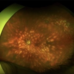 Birdshot Retinochoroidopathy
Jun 18 2025 by César Adrián Gómez Valdivia, MD
Fundus photograph of a 86 YO female patient diagnosed with Birdshot Retinochoroidopathy. Characteristically multifocal cream-colored or yellow-orange, oval or round lesions that emerge from around the optic nerve can be appreciated.
Photographer: @eyemissu2
Imaging device: California ICG OPTOS
Condition/keywords: Birdshot Retinochoroidopathy
-
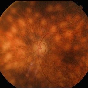 Birdshot Retinochoroidopathy
Jun 18 2025 by César Adrián Gómez Valdivia, MD
Fundus photograph of a 86 YO female patient diagnosed with Birdshot Retinochoroidopathy. Characteristically multifocal cream-colored or yellow-orange, oval or round lesions that emerge from around the optic nerve can be appreciated.
Photographer: @eyemissu2
Imaging device: TOPCON TRC-50DX
Condition/keywords: Birdshot Retinochoroidopathy
-
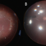 Bleb Migration With FAX
Mar 25 2025 by Robert Andrew Sisk, MD, FACS, FASRS
Color stills from surgical video after subretinal delivery of gene augmentation therapy with voretigene neparvovec-rzyl A) before and B) after fluid-air exchange (FAX). The blebs were between 0.5- and 1-disc diameters from the fovea. After FAX, they gradually extended beneath the fovea and eventually merged. This spared the fovea the trauma from the injection pressure of subretinal injection while allowing treatment to the area.
Imaging device: Leica Proveo 8
Condition/keywords: Fluid-Air Exchange, Gene Therapy, genetic disorder, genetics, Subretinal Injection
-
 Branch Retinal Vein Occlusion
Aug 30 2013 by James B. Soque, CRA, OCT-C, COA, FOPS
77-year-old female with BRVO and shunt vessel formation of the right eye.
Photographer: James B Soque, CRA, COA
Imaging device: Topcon TRC 50DX with OIS 5 MP Camera, and (MERGE) Software program
Condition/keywords: branch retinal vein occlusion (BRVO)
-
 BRVO and VMT Vitreo Macular Traction, Color Photo
Apr 18 2013 by James B. Soque, CRA, OCT-C, COA, FOPS
Color Photograph, 50 degrees, mag 2X of 79-year-old white female, VA sc 20/40, with BRVO OS, and VMT OS, diagnosed on exam and SD OCT. See accompanying RF and FA photos reveal BRVO IT OS, and SD OCT reveals VMT OS.
Photographer: James B. Soque, CRA, COA, Island Retina, Shirley, NY
Imaging device: Topcon TRC-50DX with MERGE software
Condition/keywords: branch retinal vein occlusion (BRVO), vitreomacular traction (VMT)
-
 BRVO and VMT Vitreo Macular Traction, FA Early Phase
Apr 18 2013 by James B. Soque, CRA, OCT-C, COA, FOPS
Early FA photo, 50 degrees, mag 2X of 79-year old white female, VA sc 20/40, with BRVO OS, and VMT OS, diagnosed on exam and SD OCT. See accompanying FC and RF photos reveal BRVO IT OS, and SD OCT reveal VMT OS.
Photographer: James B. Soque, CRA, COA, Island Retina. Shirley, NY
Imaging device: Topcon TRC-50DX with MERGE software
Condition/keywords: branch retinal vein occlusion (BRVO), vitreomacular traction (VMT)
-
 BRVO and VMT Vitreo Macular Traction, Red Free Photo
Apr 18 2013 by James B. Soque, CRA, OCT-C, COA, FOPS
Red Free Photo, 50 degrees, mag 2X of 79-year-old white female, VA sc 20/40, with BRVO OS, and VMT OS, diagnosed on exam and SD OCT. See accompanying FC and FA photos reveal BRVO IT OS, and SD OCT reveal VMT OS.
Photographer: James B. Soque, CRA, COA, Island Retina, Shirley, NY
Imaging device: Topcon TRC-50DX with MERGE software
Condition/keywords: branch retinal vein occlusion (BRVO), vitreomacular traction (VMT)
-
 BRVO, FA, Hemorrhage, Diabetic
Mar 13 2014 by James B. Soque, CRA, OCT-C, COA, FOPS
51-year-old white male, diabetes, and with BRVO left eye, early phase 36 seconds. Flame heme from ON, showing microaneurysims, and fine capillary detail of this FA.
Photographer: James B Soque, CRA COA
Imaging device: Topcon TRC 50DX with MERGE software
Condition/keywords: branch retinal vein occlusion (BRVO), diabetes, FA early phase, microaneurysms
 Loading…