Search results (8 results)
-
 Extra-scleral Extension of Choroidal Melanoma
Dec 23 2021 by Jessica Norkus
89-year-old female with extra-scleral extension of choroidal metastatic melanoma. Patient hadn't been seen by any eye doctor in 3 years prior to this visit. Noticed scleral darkening about 6 months ago, with vision loss noted for about 4-5 months. Presented with LP vision. Emergent MRI of brain/orbit showed no extension beyond what is seen at slit lamp. CT C/A/P w/ contrast ordered and found 2 hepatic lesions, concerning for potential mets. Patient referred to medical oncology.
Photographer: Jessica Norkus, COA, OSC
Imaging device: Topcon TRC 50DX
Condition/keywords: external photography, extrascleral extension, metastatic cancer, metastatic lesion
-
 Ocular Hypotony Due to Leaking Bleb
Apr 1 2019 by Anfisa Ayalon, MD
81-year-old male who had trabeculectomy in his right eye 4 years ago, presented to the emergency room with complains of decreased vision in that eye for two months. Slit-lamp examination showed cystic bleb with leakage, intraocular pressure was 0 MMHg. Fundus examination showed hypotony maculopathy, peripheral choroidal detachments, multiple chorioretinal folds with subretinal fluid.
Photographer: Anfisa Ayalon, MD., Meir Medical Center, Kfar Saba, Israel.
Imaging device: California, Optos 200 DTX
Condition/keywords: choroidal detachment, hypotonous retinopathy, hypotony maculopathy
-
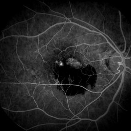 Subretinal Hemorrhage Due to SRNVM, Fluorescein Angiogram Photograph
Dec 1 2016 by James B. Soque, CRA, OCT-C, COA, FOPS
89-year-old white male with NVAMD and new subretinal hemorrhage, fluorescein angiogram, early phase, of the right eye. Currently receiving anti VEGF treatment.
Photographer: James Soque, CRA, OCT-C, COA, Island Retina, Shirley, NY
Imaging device: Topcon TRC 50 DX, with MERGE software
Condition/keywords: hemorrhage, Hot spot, neovascular age-related macular degeneration (AMD), subretinal hemorrhage, subretinal blood, wet age-related macular degeneration (wet AMD)
-
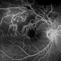 Branch Retinal Vein Occlusion- Fluorescein Angiogram, Montage
Apr 15 2016 by James B. Soque, CRA, OCT-C, COA, FOPS
A fluorescein angiogram of an 80-year-old white female with a superotemporal branch retinal vein occlusion, and retinal edema of the right eye. Currently receiving Lucentis 0.5 injection therapy.
Photographer: James Soque, CRA OCT-C COA, Island Retina, Shirley, NY
Imaging device: Topcon TRC, MERGE Imaging Software V. 11.2.0
Condition/keywords: branch retinal vein occlusion (BRVO), montage, non-perfused branch retinal vein occlusion (BRVO)
-
 Choroidal Melanoma
Jan 8 2016 by Jared Watson
42-year-old white female, S/P I-125 plaque brachytherapy.
Photographer: Jared Watson COT/CRA
Imaging device: Topcon 50EX, OIS-Merge
-
 Hemi Vein Occlusion, Fluorescein Angiogram, Montage
Dec 17 2015 by James B. Soque, CRA, OCT-C, COA, FOPS
74-year-old woman, with recurrent superior hemi vein occlusion, montage image of fluorescein angiogram left eye. Currently receiving Lucentis injections OS.
Photographer: James Soque, CRA COA
Imaging device: Topcon RC 50 DX Fundus Camera with MERGE Winstation Software for Fluorescen Angiography
Condition/keywords: montage, occlusion of retinal vein, superior arcade
-
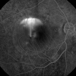 CSCR Mushroom Cloud
Feb 23 2015 by James J. Bedrick, MD
Late transit FA of a large active sub-foveal CSCR leak. You may view this pair in stereo to appreciate the plume of leakage within this large serous RD of the macula. This patient presented with a BCVA of 20/200 and fluorescein and historic evidence of prior episodes of leakage. After discussion of known treatment options including observation, he elected to be treated initially with oral rifampin and BCVA improved to 20/40 with persistent metamorphosis and a shallower persistent macular detachment over several visits. Rifampin was discontinued and he then received sub-threshold micro-pulse laser photocoagulation with an 810 diode which resulted in the patient reporting full restoration of his vision subjectively within a month. He failed to keep his follow-up appointment.
Photographer: Diana Bodnar, COT
Imaging device: Topcon 50X with Merge capture station
Condition/keywords: CSCR subfoveal leak
-
 Neovascular ARMD With Subretinal Hemorrhage, Red-Free Photos - Stereo
Nov 26 2014 by James B. Soque, CRA, OCT-C, COA, FOPS
Stereo FC, RF and FA of a 77-year-old white female with visual acuity CC 20/200-3, with left eye neovascular ARMD, drusen, and subretinal hemorrhage with hard exudates temporally. Peripheral retina reveals cobblestone degeneration.
Photographer: James Soque, CRA, COA, Island Retina, Shirley, NY
Imaging device: Topcon TRC 50 EX, with MERGE software and OIS 5 MP digital Camera
Condition/keywords: neovascular age-related macular degeneration (AMD), red-free, stereo pair
 Loading…