Search results (87 results)
-
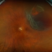
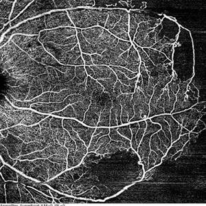
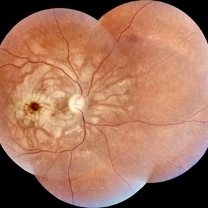 Purtscher-like Retinopathy in Preeclampsia
Jun 28 2025 by Sriharanathan Poopalaratnam, MD,FRCS
A 24-year-old female, 6 weeks post-emergency cesarean section (LSCS), with a history of pregnancy-induced hypertension (PIH), presented with acute, profound, bilateral painless vision loss of 2 days’ duration
Photographer: Yattiwarra
Condition/keywords: preeclampsia, Purtscher like
-
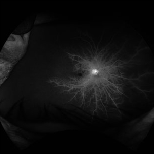
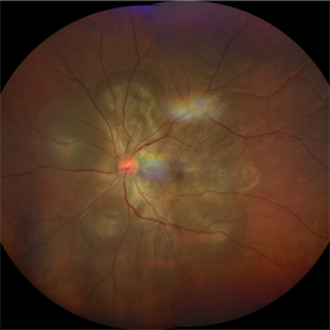
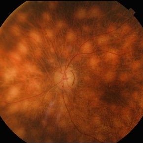 Birdshot Retinochoroidopathy
Jun 18 2025 by César Adrián Gómez Valdivia, MD
Fundus photograph of a 86 YO female patient diagnosed with Birdshot Retinochoroidopathy. Characteristically multifocal cream-colored or yellow-orange, oval or round lesions that emerge from around the optic nerve can be appreciated.
Photographer: @eyemissu2
Imaging device: TOPCON TRC-50DX
Condition/keywords: Birdshot Retinochoroidopathy
-
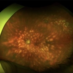 Birdshot Retinochoroidopathy
Jun 18 2025 by César Adrián Gómez Valdivia, MD
Fundus photograph of a 86 YO female patient diagnosed with Birdshot Retinochoroidopathy. Characteristically multifocal cream-colored or yellow-orange, oval or round lesions that emerge from around the optic nerve can be appreciated.
Photographer: @eyemissu2
Imaging device: California ICG OPTOS
Condition/keywords: Birdshot Retinochoroidopathy
-
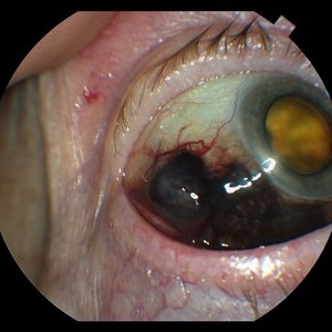
 Bleb Migration With FAX
Mar 25 2025 by Robert Andrew Sisk, MD, FACS, FASRS
Color stills from surgical video after subretinal delivery of gene augmentation therapy with voretigene neparvovec-rzyl A) before and B) after fluid-air exchange (FAX). The blebs were between 0.5- and 1-disc diameters from the fovea. After FAX, they gradually extended beneath the fovea and eventually merged. This spared the fovea the trauma from the injection pressure of subretinal injection while allowing treatment to the area.
Imaging device: Leica Proveo 8
Condition/keywords: Fluid-Air Exchange, Gene Therapy, genetic disorder, genetics, Subretinal Injection
-
 Retinal Detachment with Horseshoe Retinal Tear
Feb 17 2025 by Kimberly Wakester
Optomap RGB image of a 62-year-old woman with a retinal detachment with a horseshoe retinal tear in the left eye. Patient had emergent surgery same day. She is doing well post operatively. Will continue follow up care as directed.
Photographer: Kimberly Wakester, COA
Imaging device: Optos California
Condition/keywords: horseshoe tear, retinal detachment
-
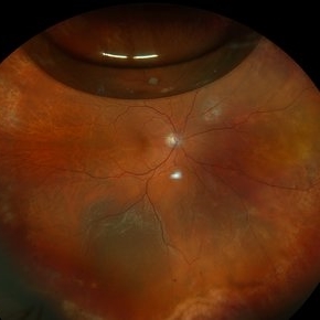
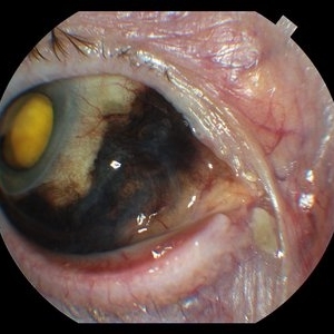
 Mac-on Retinal Detachment (Barely!)
Feb 6 2025 by Virginia Gebhart
FAF of 46 year old male with a mac-on retinal detachment from 1:00 to 6:00 with a single break at 3:00. Pt scheduled for emergent PPV/Laser/GFE
Photographer: Virginia Gebhart, Retina Consultants of Carolina
Imaging device: Optos California
Condition/keywords: autofluorescence imaging, retinal detachment
-
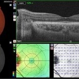
 Treatment of Ocular Ischemic Syndrome with Hyperbaric Oxygen Therapy
Dec 2 2024 by Catherine S Kang
A 66-year-old female with past medical history significant for hypertension and ocular ischemic syndrome. She presented in emergency department (ED) reporting eye pain and blurred vision in both eyes since earlier that morning. On examination, best corrected visual acuity in each eye was counting fingers (20cm). Further investigation was performed and fluorescein angiography revealed a delay in choroidal filling. The patient has been followed for ocular ischemic syndrome since the onset of the condition and hyperbaric oxygen therapy was promptly initiated. Final best corrected visual acuity was 20/150 and macula developed atrophy.
Photographer: Catherine Kang
Condition/keywords: hyperbaric oxygen therapy, ocular ischemic syndrome
-
 Recurrent Retinal Detachment with Single Break
Nov 2 2024 by Virginia Gebhart
84 year old male with recurrent detachment s/p PPV/RD repair 2 weeks ago. Retinotomy is opened and appears to be the source of the fluid. Pt scheduled for emergency repair with scleral buckle.
Photographer: Virginia Gebhart
Imaging device: Optos California
-
 Macular Star
Sep 8 2024 by Cesar Augusto Rocha Rojas, MD
Fundus photograph of a 27-year-old male with hypertensive emergency secondary to chronic kidney disease.
Photographer: Cesar Augusto Rocha Rojas, Hospital General de Zona #20, Instituto Mexicano del Seguro Social (IMSS)
Imaging device: Smartphone, Pan Retinal 2.2 Lens
Condition/keywords: macular star
-
 Hypertensive Retinopathy
Sep 8 2024 by Cesar Augusto Rocha Rojas, MD
Fundus photograph of a 27-year-old male with hypertensive emergency secondary to chronic kidney disease.
Photographer: Cesar Augusto Rocha Rojas, Hospital General de Zona #20, Instituto Mexicano del Seguro Social
Imaging device: Smartphone, Pan Retinal 2.2 Lens
Condition/keywords: hypertensive retinopathy
-
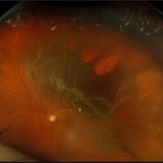 Rhegmatogenous Macula Off Retinal Detachment with Multiple Breaks
May 29 2024 by Alexis Singstock
Ultra widefield fundus photograph of a 66 year old male with rhegmatogenous macula off retinal detachment with multiple breaks. Patient presented emergently for a curtain/veil in inferonasal visual field. Patient reports the curtain/veil in left eye started about 1 week prior, yet denied seeing flashes and floaters. Patient's vision was hand motion. Dr. Edward Korot examined the patient and scheduled him for a scleral buckle along with pars plana vitrectomy surgery.
Photographer: Alexis Singstock, Retina Specialists of Michigan
Imaging device: Optos California
Condition/keywords: fundus photography, left eye, macula off retinal detachment, OPTOS CALIFORNIA, pars plana vitrectomy (PPV), rhegmatogenous retinal detachment, scleral buckle, ULTRA WIDE FIELD
-
 Situs-Inversus-Left-eye
Mar 29 2023 by Nizamuddin HM Shaik, MD, FRCS
Situs inversus of the optic disc is a rare, usually bilateral, congenital embryological abnormality associated with high myopia, optic disc coloboma or tilted optic disc. Our patient, 24 years old lady without these conditions presented with bilateral situs inversus. Her BCVA OD 0.4 and OS 0.5. It is characterized by emergence of the retinal vessels in an anomalous direction with dysversion of the optic disc.
Photographer: Mahmoud A Abdelmaguid
Condition/keywords: Nasalization of temporal retinal vessels
-
 Situs-Inversus-OD
Mar 29 2023 by Nizamuddin HM Shaik, MD, FRCS
Situs inversus of the optic disc is a rare, usually bilateral, congenital embryological abnormality associated with high myopia, optic disc coloboma or tilted optic disc. Our patient, 24 years old lady without these conditions presented with bilateral situs inversus. Her BCVA OD 0.4 and OS 0.5. It is characterized by emergence of the retinal vessels in an anomalous direction with dysversion of the optic disc.
Photographer: Mahmoud A Abdelmaguid
Condition/keywords: Nasalization of temporal vessels
-
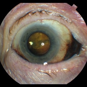
 Central retinal artery occlusion
Nov 30 2022 by Ethan K Sobol, MD
A central retinal artery occlusion with cilioretinal artery sparing, imaged using a Volk Panretinal 2.2 and an iPhone camera in the emergency department.
Photographer: Jared Raabe, MD, Emory University Hospital
Imaging device: IPhone 13 Pro
Condition/keywords: central retinal artery occlusion (CRAO)
-
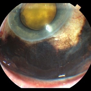
 Silicon Oil in CT brain
Aug 5 2022 by Jesus Lozano, MD
An 65-year-old woman was taken to the Emergency Department after a fall. CT brain imaging demonstrated a well-defined, homogenous, hyperdense mass in the posterior segment of the right eye. Detailed history revealed previous vitreoretinal procedures for multiple retinal detachments. Ophthalmological examination confirmed presence of silicone oil in this eye.
Photographer: Dr. Jesus Lozano Gutierrez
Condition/keywords: retina surgery
-
 Vogt-Koyanagi-Harada Disease
Apr 24 2022 by Aniruddha K Agarwal, MD
A 38-year-old woman of Asian descent with no ophthalmological or systemic history presented to the emergency eye clinic with a 1-week complaint of headache and bilateral vision loss. Funduscopy revealed bilateral serous neurosensory detachments. The presence of lymphocytosis in cerebrospinal fluid and mild acute sensorineural hearing loss confirmed the diagnosis of uveomeningoencephalitic syndrome (Vogt-Koyanagi-Harada disease).
Photographer: Mercedes SERRADOR, MD, PhD and Beatriz VENTAS, MD
Imaging device: Zeiss Clarus fundus camera
Condition/keywords: IUSG, panuveitis, Vogt-Koyanagi-Harada
-
In vivo disseminated central retinal artery embolism
Mar 22 2022 by Argyrios Chronopoulos, MD, FMH, MRCP
Central retinal artery occlusion (CRAO) is a retinal stroke and although ophthalmological treatment is controversial, emergency cardiovascular and neurological evaluation is indicated because patients with CRAO are prone to cerebral stroke within the first 7 to 30 days.Emboli are not always present or visible. This case example may assist those involved in diagnosis and management of CRAO patients including general practitioners, internists, emergency physicians, neurologists, cardiologists as well as ophthalmologists.
Photographer: A. Chronopoulos
Imaging device: Haag Streit Slitlamp with mounted Camera/Video Module
Condition/keywords: amaurosis fugax, carotid stenosis, central retinal artery occlusion (CRAO), retinal embolis
-
 Extra-scleral Extension of Choroidal Melanoma
Dec 23 2021 by Jessica Norkus
89-year-old female with extra-scleral extension of choroidal metastatic melanoma. Patient hadn't been seen by any eye doctor in 3 years prior to this visit. Noticed scleral darkening about 6 months ago, with vision loss noted for about 4-5 months. Presented with LP vision. Emergent MRI of brain/orbit showed no extension beyond what is seen at slit lamp. CT C/A/P w/ contrast ordered and found 2 hepatic lesions, concerning for potential mets. Patient referred to medical oncology.
Photographer: Jessica Norkus, COA, OSC
Imaging device: Topcon TRC 50DX
Condition/keywords: external photography, extrascleral extension, metastatic cancer, metastatic lesion
-
 Extra-scleral Extension of Choroidal Melanoma
Dec 23 2021 by Jessica Norkus
89-year-old female with extra-scleral extension of choroidal metastatic melanoma. Patient hadn't been seen by any eye doctor in 3 years prior to this visit. Noticed scleral darkening about 6 months ago, with vision loss noted for about 4-5 months. Presented with LP vision. Emergent MRI of brain/orbit showed no extension beyond what is seen at slit lamp. CT C/A/P w/ contrast ordered and found 2 hepatic lesions, concerning for potential mets. Patient referred to medical oncology.
Photographer: Jessica Norkus, COA, OSC
Imaging device: Topcon TRC 50DX
Condition/keywords: extrascleral extension, metastatic cancer, metastatic lesion
-
 Extra-scleral Extension of Choroidal Melanoma
Dec 23 2021 by Jessica Norkus
89-year-old female with extra-scleral extension of choroidal metastatic melanoma. Patient hadn't been seen by any eye doctor in 3 years prior to this visit. Noticed scleral darkening about 6 months ago, with vision loss noted for about 4-5 months. Presented with LP vision. Emergent MRI of brain/orbit showed no extension beyond what is seen at slit lamp. CT C/A/P w/ contrast ordered and found 2 hepatic lesions, concerning for potential mets. Patient referred to medical oncology.
Photographer: Jessica Norkus, COA, OSC
Imaging device: Topcon TRC 50DX
Condition/keywords: external photography, extrascleral extension, metastatic cancer, metastatic lesion
-
 Extra-scleral Extension of Choroidal Melanoma
Dec 23 2021 by Jessica Norkus
89-yea- old female with extra-scleral extension of choroidal metastatic melanoma. Patient hadn't been seen by any eye doctor in 3 years prior to this visit. Noticed sclera darkening about 6 months ago, with vision loss noted for about 4-5 months. Presented with LP vision. Emergent MRI of brain/orbit showed no extension beyond what is seen at slit lamp. CT C/A/P w/ contrast ordered and found 2 hepatic lesions, concerning for potential mets. Patient referred to medical oncology.
Photographer: Jessica Norkus, COA, OSC
Imaging device: Topcon TRC 50DX
Condition/keywords: external photography, extrascleral extension, metastatic cancer, metastatic lesion
-
 Extra-scleral Extension of Choroidal Melanoma
Dec 23 2021 by Jessica Norkus
89-year-old female with extra-scleral extension of choroidal metastatic melanoma. Patient hadn't been seen by any eye doctor in 3 years prior to this visit. Noticed sclera darkening about 6 months ago, with vision loss noted for about 4-5 months. Presented with LP vision. Emergent MRI of brain/orbit showed no extension beyond what is seen at slit lamp. CT C/A/P w/ contrast ordered and found 2 hepatic lesions, concerning for potential mets. Patient referred to medical oncology.
Photographer: Jessica Norkus, COA, OSC
Imaging device: Topcon TRC 50DX
Condition/keywords: extrascleral extension, metastatic cancer, metastatic lesion
-
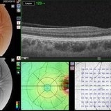
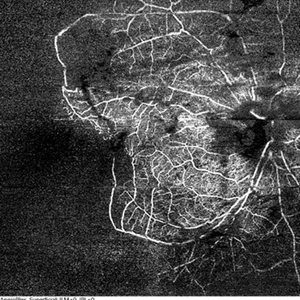
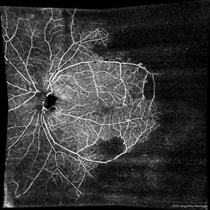 Ischemic CRVO
Jun 23 2021 by Eduardo Torres-Porras, MD
Montage 8x8 mm OCT angiography of the left eye of a 36-year-old male who had an ischemic CRVO following a hypertensive emergency secondary to consumption of high doses of cocaine. Areas of non perfusion can be seen within the posterior pole which extends temporally to the mid-periphery.
Photographer: Eduardo Torres-Porras, Provissia, Laser y Ultrasonido Ocular
Imaging device: Cirrus 600, Carl Zeiss
Condition/keywords: central retinal vein occlusion (CRVO), ischemic CRVO, optical coherence tomography (OCT), ultra-wide field imaging
-
 Ischemic CRVO
Jun 23 2021 by Eduardo Torres-Porras, MD
HD 8x8 mm OCT angiography of a 36-year-old male who had an ischemic CRVO following a hypertensive emergency secondary to consumption of high doses of cocaine. The posterior pole has areas of non perfusion.
Photographer: Eduardo Torres-Porras, Provissia, Laser y Ultrasonido Ocular
Imaging device: Cirrus 600, Carl Zeiss
Condition/keywords: central retinal vein occlusion (CRVO), ischemic CRVO, optical coherence tomography (OCT), ultra-wide field imaging
-
 Ischemic CRVO
Jun 23 2021 by Eduardo Torres-Porras, MD
12x12 mm OCT angiogram of the right eye of a 36-year-old male who had an ischemic CRVO following a hypertensive emergency secondary to consumption of high doses of cocaine. Areas of non perfusion are found within the posterior pole and ischemia extends temporally to the mid-periphery.
Photographer: Eduardo Torres-Porras, Provissia, Laser y Ultrasonido Ocular
Imaging device: Cirrus 600, Carl Zeiss
Condition/keywords: central retinal vein occlusion (CRVO), ischemic CRVO, optical coherence tomography (OCT), ultra-wide field imaging
 Loading…