Discover images
-
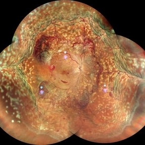
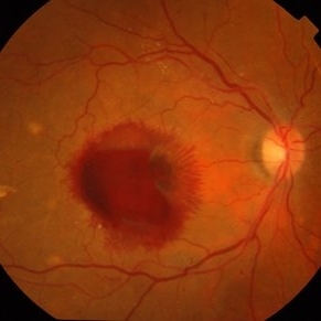
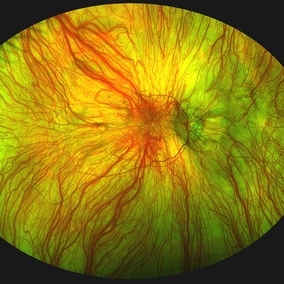
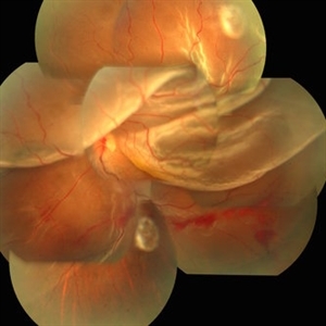 Globe Perforation With Retinal Detachment
Feb 7 2017 by Manish Nagpal, MD, FRCS (UK), FASRS
Patient presenting with globe perforation with a penetration seen at below the inferior arcade with some scattered hemorrhage and a retinal detachment.
Photographer: Rakesh Juneja
Condition/keywords: globe perforation
-
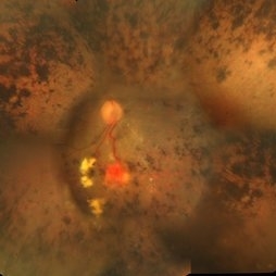
 Total Rhegmatogenous Retinal Detachment With Severe PVR
May 27 2015 by Darin R. Goldman, MD
63-year-old pseudophakic male with hand motion vision in the left eye due to a total retinal detachment with severe proliferative vitreoretinopathy.
Condition/keywords: proliferative vitreoretinopathy (PVR), retinal tear
-
 Wide-Field-OCT-montage
Jan 8 2018 by Netan Choudhry, MD, FRCS(C) FASRS
This is an SD-OCT montage image of a 55 year old male with optic neuropathy representing a wide-field OCT spanning 130 degrees.
Photographer: John Golding, Vitreous Retina Macula Specialists of Toronto
Imaging device: Heidelberg Spectralis OCT system
Condition/keywords: wide angle imaging
-
 Coats' Disease
Apr 27 2018 by Brenda Fallas
3-year-old boy with unilateral Coats' Disease fundus photo.
Photographer: Brenda Fallas, Bascom Palmer Eye Institute, Miami, FL
Imaging device: Retcam III 130 degree lens
Condition/keywords: Coats' disease, color fundus photograph, retinal telangiectasia
-
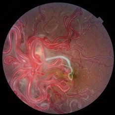 Wyburn Mason
Jun 23 2018 by Caesar K. Luo, MD, FASRS
Fundus photograph of a 16-year-old female patient with poor vision in one eye demonstrates racemose angiomatosis as seen in Wyburn-Mason Syndrome.
Photographer: Joseph Trabucco, Progressive Vision Institute, Allentown, PA
Condition/keywords: Wyburn-Mason
-
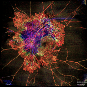 Flame of the Forest
Apr 9 2020 by Daraius N Shroff, MS FMRF FRCS
A 54-year-old man with DM for 15 years. The left eye had a visual acuity of 20/40. Wide field swept source OCTA revealed branching out central neovascular trunk vessels from the disc with terminal loops, along with exuberant proliferation of irregular small-calibre fine new vessels. The patient underwent OCTA guided pan retinal photocoagulation.
Photographer: Anuj Choudhary, Shroff Eye Centre, New Delhi
Imaging device: Zeiss Plex Elite 9000
Condition/keywords: proliferative diabetic retinopathy (PDR)
-
 Proliferative Diabetic Retinopathy with Choroidal Effusion Status Post PRP
Dec 15 2020 by Manish Nagpal, MD, FRCS (UK), FASRS
A 17-year-old juvenile diabetic patient came to us with extensive neovascular proliferations and PRP done a week back and had developed 360 degree choroidal effusion as seen in this wide field montage image
Photographer: Sham Talati, Retina Fellow , Retina Foundation, Ahmedabad, India
Imaging device: Mirante CSLO
Condition/keywords: choroidal effusion, diabetic retinopathy, proliferative diabetic retinopathy (PDR)
-
 Retinitis Pigmentosa With Hemangioma CF
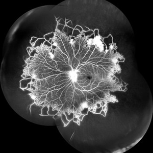
Dec 15 2016 by Manish Nagpal, MD, FRCS (UK), FASRS
Fluorescein angiography OS of a patient having retinitis pigmentosa with a hemangioma inferiorly.
Condition/keywords: hemangioma, retinitis pigmentosa
-
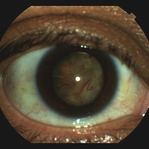
 Retinoblastoma
Apr 27 2018 by Brenda Fallas
2-year-old boy with stage D+ retinoblastoma of the right eye.
Photographer: Brenda Fallas, Bascom Palmer Eye Institute, Miami, FL
Imaging device: RETCAM III 130 degree lens montage
Condition/keywords: tumor, tumor seeding
-
 Valsalva Retinopathy
Jan 26 2017 by JEFFERSON R SOUSA, Tecg.º (Biomedical Systems Technology)
Male patient, 23-years-old, with low visual acuity in the right eye. In the ocular examination of the retinography, intense subhyaloidal hemorrhage. 2 minutes after laser application.
Photographer: JEFFERSON R SOUSA - Suel Abujamra Institute - São Paulo - Brazil
Imaging device: Topcon TRC-50 DX, Imaginet, 35 degree field. Flash 36 / Mosaic with four images.
Condition/keywords: subhyaloid hemorrhage, valsalva retinopathy
-
 Coats' Disease FA
Apr 27 2018 by Brenda Fallas
3-year-old boy with unilateral Coats' Disease FA photo.
Photographer: Brenda Fallas, Bascom Palmer Eye Institute, Miami, FL
Imaging device: Retcam III 130 degree lens
Condition/keywords: Coats' disease, FA early phase, fluorescein angiogram (FA), retinal telangiectasia
-
 Macroaneurysm
Apr 1 2017 by Manish Nagpal, MD, FRCS (UK), FASRS
Case of a ruptured macroaneurysm with subhyaloid and subretinal blood.
Photographer: Avijit Vishnoi
Condition/keywords: macroaneurysm, ruptured macroaneurysm
-
 Post Traumatic Optic Nerve Head Avulsion
Nov 18 2017 by Vishal Agrawal, MD, FRCS,FACS,FASRS
Right eye fundus picture of a 24-year-old male patient who suffered blunt trauma 7 days back with a wooden stick . He presented with NLP vision with a non reacting dilated pupil. Fundus montage picture shows ONH avulsion,CRAO,peripapillary resolving hemorrhages and cicatricial tissue at the edge.
Photographer: Vishal Agrawal, MD, SMS Medical College, Jaipur, India
Imaging device: Zeiss 524
Condition/keywords: avulsion, central retinal artery occlusion (CRAO)
-
 Tractional Retinal Detachment
Dec 4 2019 by Janet Brazil
Fundus photograph of a 32-year-old female with severe end-stage diabetic tractional retinal detachment.
Photographer: Janet Atkinson, Eye Associates of New Mexico, Albuquerque, NM
Imaging device: Topcon TRC- 50EX
Condition/keywords: diabetes, proliferative diabetic retinopathy (PDR), tractional retinal detachment
-
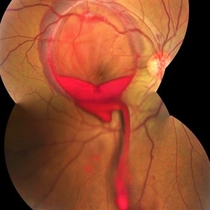
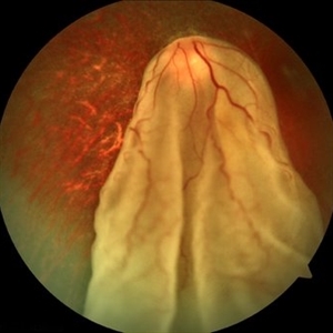 When the Curtain Falls
Jun 12 2021 by Shyamal K Dwivedi, MD
52-year-old male presented with sudden painless vision drop. Giant retinal tear discovered which was about 270 degrees anchored at the disc. Title courtesy: Dr.Aditya Sudhalkar
Photographer: Dr.Aditya Sudhalkar
Imaging device: Zeiss
Condition/keywords: giant retinal tear
-
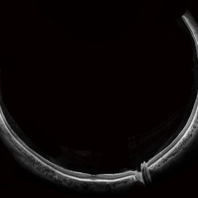
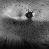
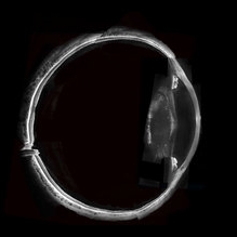 Whole Eye OCT
Jan 4 2019 by Netan Choudhry, MD, FRCS(C) FASRS
Swept-Source OCT montage of a 45-year-old male with Alports disease and posterior subcapsular cataract.
Photographer: John Golding BA, Vitreous Retina Macula Specialists of Toronto
Imaging device: Topcon DRI Triton
Condition/keywords: Alports disease, optical coherence tomography (OCT), swept source
-
 Albinotic Fundus
Jan 24 2024 by Poornachandra B, MS, FVRS
Fundus photo of a 30 year old male with Ocular albinism. Hypopigmented fundus with very evident choroidal vessels.
Photographer: Dr Poornachandra B
Condition/keywords: ocular albinism
-
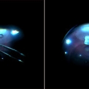 Foreign Body SS
Feb 14 2015 by Shlomit Schaal, MD, PhD, MHCM
A four millimeter metal foreign body removed surgically using foreign body forceps. The macula is protected by PFCL heavy fluid. There is a traumatic retinal detachment inferiorly (top of photo, surgeon's view).
Photographer: Shlomit Schaal, MD, PhD
Condition/keywords: intraocular foreign body
-
 Massive Subretinal Hemorrhage With Near Total Retina Detachment
Nov 27 2013 by David W. Faber, MD
Fundus photo of an 71-year-old male with massive subretinal hemorrhage. Had been given 6 week Avastin treatments. Was put on coumadin for 6 weeks following knee surgery.
Photographer: Donna Knight, Rocky Mountain Retina Consultants, Salt Lake City, Utah
-
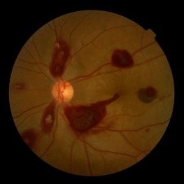 Ocular Manifestation of Acute Leukemia
Sep 8 2012 by Hamid Ahmadieh, MD
Color fundus photograph of a 26-year-old man with acute leukemia.
Photographer: Hamid Ahmadieh, MD, Ophthalmic Research Center, Labbafinejad Medical Center, Shahid Beheshti University of Medical Sciences , Tehran
Imaging device: Topcon Fundus Camera
Condition/keywords: acute leukemia, white centered retinal hemorrhage (Roth Spot)
-
 Phoenix
Feb 21 2024 by Sayena . Jabbehdari, MD, MPH, MBA
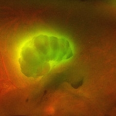
A 60-year-old Caucasian female presented with reduced night vision and constricted visual fields. The fundus exam revealed pigmentary changes in the peripheral retina. Fundus autofluorescence depicted the schematic appearance of a Phoenix , with the hypo-autofluorescence corresponding to the head and wings of the phoenix. Genetic testing was positive for a heterozygous RHO mutation
Photographer: Sayena Jabbehdari MD MPH
Condition/keywords: retinitis pigmentosa
-
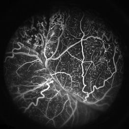
 Vascular Non Perfusion in Takayasu Arteritis
Feb 6 2024 by SHILPI H NARNAWARE, ICO ( Retina) , FAICO ( Vitreo-Retina)
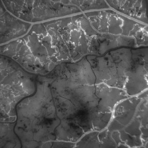
A case of 16 year-old female with combined RD in RE. Fundus examination & FFA revealed 360 degrees non-perfusion in periphery in non-symptomatic eye.
Photographer: Shilpi Narnaware, Sarakshi Netralaya , Nagpur, Maharashtra , India
Imaging device: Mirante ( by Nidek)
Condition/keywords: CNP areas, takayasu arteritis
-
 Acute Posterior Multifocal Placoid Pigment Epitheliopathy
Feb 20 2024 by Soobien Lee
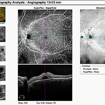
12x12mm OCT Angiography of a 20-year-old caucasian female with viral prodrome and vision loss OS>OD secondary to Acute Posterior Multifocal Placoid Pigment Epitheliopathy (APPME). Imaging shows multifocal flow voids.
Photographer: Kim Seay, Elman Retina Group
Imaging device: 12x12mm OCT-Angiography
Condition/keywords: acute posterior multifocal placoid pigment epitheliopathy (APMPPE), bacillary layer detachment, OCT, OCT Angiography, Uveitis, white dot syndrome
-
 Aurora Borealis in Retina
Apr 25 2025 by Poornachandra B, MS, FVRS
Fundus picture of 54 year old male with proliferative diabetic retinopathy with fluorescent blood clot in vitreous cavity.
Photographer: Mr Dhikshith
Imaging device: Optos daytona
Condition/keywords: blood, proliferative diabetic retinopathy (PDR)
-
 Branch Retinal Vein Occulsion
Sep 14 2012 by Michael P. Kelly, FOPS
This is a high-magnification fluorescein angiogram image of an area distal to the point of a branch retinal vein occlusion.
Photographer: Michael P. Kelly, FOPS, Director, Duke Eye Center Labs, Duke University Hospital
Condition/keywords: branch retinal vein occlusion (BRVO), high magnification, ischemia
 Loading…