Discover images
-
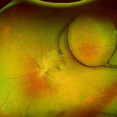
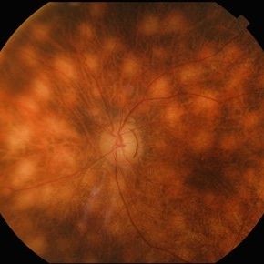
 Retinal Detachment with Macular Hole
Nov 10 2025 by Korey Starkey
A 58 year-old female presented with re-detached retina through macular hole. Planned for surgical intervention.
Photographer: Korey Starkey
Imaging device: Optos
Condition/keywords: gas bubble, intermediate uveitis, macular hole, pars plana vitrectomy (PPV), retinal detachment, scleral buckle, traction detachment
-
 Posterior Dislocated Intraocular Lens
Oct 23 2025 by Aditya S Kelkar, MS, FRCS, FASRS,FRCOphth
Fundus photograph of a 53-year-old man with a posteriorly dislocated intraocular lens near the posterior pole.
Photographer: Dr Tejal Rao, National Institute of Ophthalmology, Pune, India
Imaging device: Optos Daytona
Condition/keywords: dislocated intraocular lens (IOL), IOL drop
-
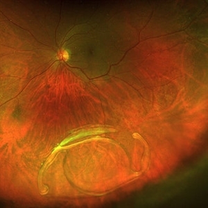
 RD with GRT with Dislocated Cataractous Lens
Nov 9 2025 by SHILPI H NARNAWARE, ICO ( Retina) , FAICO ( Vitreo-Retina)
Young high myope male presented with DOV since 6 months. Examination revealed , Subluxated cataractous lens with RD with PVR with > 270 degrees GRT
Photographer: Shilpi Narnaware, Sarakshi Netralaya , Nagpur, Maharashtra , India
Imaging device: Mirante ( by Nidek)
Condition/keywords: PVR, retinal detachment
-
 Proliferative Diabetic Retinopathy
Nov 13 2025 by DR APOORVA JADHAV, MBBS , DNB
Wild Field OCTA of 61 year old male showing us NVD, NVE, CNP areas , macular ischaemia.
Photographer: Dr Apoorva Jadhav
Imaging device: Intalight Dream OCT
Condition/keywords: neovascularization of the disc (NVD), NVE, PDR
-
 Anterior Migration of the Dex Implant
Nov 22 2025 by Gabriel Costa Andrade, PhD
Anterior segment photograph of a Ozurdex implant in a 53-year-old man with macular edema due to intermediate uveitis.
Photographer: Gabriel Andrade
Condition/keywords: Ozurdex implant
-
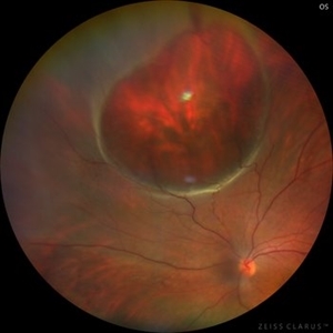
 Origami Under Silicone Oil
Nov 29 2025 by Resham Khatri
Fundus photograph of a woman presenting for vision loss having operated twice for retinal detachment.
Photographer: Resham Khatri, M&J WRIO, Ahmedabad
Condition/keywords: post-vitrectomy, re-detachment post-vitrectomy, retinal detachment
-
 IOL Drop
Dec 4 2025 by Surabhi Gupta, MS, DNB, FVRS
A 60 year old man presented with sudden dimunition of vision in right eye. His visual acuity was finger counting at 1 meter and best corrected visual acuity with +10 D was 6/9. Patient was diagnosed with spontaneous right eye IOL bag complex drop in vitreous cavity with superior HST and inferotemporal hole secondary to posterior vitreous detachment . Right eye montage color fundus photo shows rigid IOL bag complex in vitreous cavity with barraged superior HST and inferotemporal hole. Post barrage laser patient underwent pars plana vitrectomy with IOL explantation and scleral fixated IOL.
Photographer: Dr Surabhi Gupta
Imaging device: EDION FA
Condition/keywords: IOL drop
-
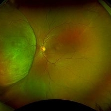
 Giant Retinal Cyst
Sep 20 2025 by JORGE SOBERANES
Fundus photograph of a 45-year-old-man with a large cyst on the nasal superior side of the retina. The patient had a history of a pneumatic retinopexy two years ago and the cyst has been there since that.
Photographer: Dr. Jorge Soberanes, Asociación para Evitar la Ceguera en México (APEC), UNAM
Condition/keywords: abnormal retina, pneumatic retinopexy, retinal cyst
-
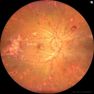 Leukemic Retinopathy and Optic Neuropathy
Aug 25 2025 by Elysse Tom, MD
Fundus photo of a 45-year-old woman with chronic myeloid leukemia.
Condition/keywords: leukemia, leukemic infiltration, Leukemic optic neuropathy
-
 Retinal Arteriovenous Malformation
Oct 7 2025 by Korey Starkey
55 year-old patient presents with retinal arteriovenous malformation in the left eye and BRVO w/retinal neovascularization. Patient is asymptomatic. No edema or treatment necessary today, signs of old RVO with MAs along inferior arcade and dot heme.
Photographer: Korey Starkey
Imaging device: Topcon
Condition/keywords: branch retinal vein occlusion (BRVO), fundus photography, inferior arcade, microaneurysms, retinal arteriovenous malformations, retinal neovascularization, Topcon
-
 Amelanotic Melanoma
Sep 19 2025 by Aditya S Kelkar, MS, FRCS, FASRS,FRCOphth
Widefield fundus photograph of a 37 year old showing a large, dome-shaped, intraocular mass involving the temporal retina. The lesion appears elevated and lacks surface pigmentation. Overlying retinal vessels are displaced and draped across the tumor surface, with surrounding retinal elevation noted. The appearance is suggestive of amelanotic variant of choroidal melanoma.
Photographer: Dr. Muskan Mangal
Imaging device: Optos Daytona
Condition/keywords: choroidal melanoma, intraocular tumor
-
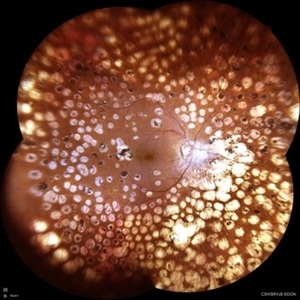
 Unexpected Sanctuary: Gas Bubble Entrapment in Morning Glory Disc
Sep 5 2025 by Danny Salgado Gómez
Fundus photograph of a 62-year-old male patient with Morning Glory syndrome in the right eye, who underwent vitrectomy, gas, and endolaser for posterior pole detachment. In the postoperative period, a gas bubble is observed within the optic disc, which persisted even after complete reabsorption of the intraocular gas.
Photographer: Dr. Danny Salgado, Retina and Vitreous Fellow, Clínica Oftalmológica del Caribe, Colombia.
Condition/keywords: gas bubble, intraocular gas, Morning Glory, Retinal Detachment, vitrectomy
-
 Diabetic Retinopathy
Aug 19 2025 by JEFFERSON R SOUSA, Tecg.º (Biomedical Systems Technology)
Female patient, 74 years old, with a history of severe photocoagulated diabetic retinopathy in both eyes.
Photographer: JEFFERSON ROCHA DE SOUSA - Retinal Department at Lens Oftalmologia, Sao Paulo-Brazil
Imaging device: EIDON fundus camera with a 110° field of view, confocal scanning technology. Widefield co-op with 4 images.
Condition/keywords: background diabetic retinopathy (BDR), Diabetic Retinopathy
-
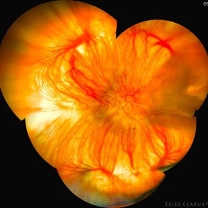 Albinotic Fundus
Aug 18 2025 by KANWALJEET HARJOT MADAN, M.S. (Ophthalmology); FAICO (Vitreous - Retina)
This is ultrawide field montage image of a 40 year-old male depicting Hypopigmented Albinotic Fundus. Arborizing network of choroidal vasculature is prominent. Multiple vortex vein ampullae near equator and posterior pole are clearly visible.
Photographer: Dr. Kanwaljeet Harjot Madan, Thind Eye Hospital, Jalandhar City (Punjab). INDIA.
Imaging device: Zeiss Fundus Camera
Condition/keywords: Albinotic Fundus, choroidal vasculature, vortex vein
-
 Presumed Congenital Toxoplasmosis
Aug 16 2025 by Vishal Agrawal, MD, FRCS,FACS,FASRS
Fundus picture of 7 a year-old boy with esotropia. OCT showed complete atrophy & disorganization of the overlying RPE and neurosensory retina.
Photographer: Dr Ayushi Gupta
Imaging device: Clarus 700
Condition/keywords: coloboma of macula, toxoplasmosis
-
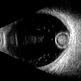
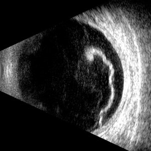 Giant Retinal Tear
Jul 5 2025 by Gustavo Uriel Fonseca Aguirre
This B-mode longitudinal ultrasound scan reveals a giant retinal tear, demonstrating a circumferential retinal flap with rolled edges extending over M-X to M-I. The vitreous shows diffuse hemorrhage and anterior-posterior traction strands inserting at the tear margins. The remaining retina appears attached without subretinal fluid.
Photographer: Gustavo U. Fonseca Aguirre, Hospital Conde de Valenciana, Ciudad de México
Condition/keywords: giant retinal tear
-
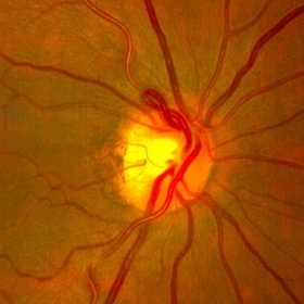 Prepapillary Vascular Loop
Jul 4 2025 by KANWALJEET HARJOT MADAN, M.S. (Ophthalmology); FAICO (Vitreous - Retina)
This is the fundus picture of right eye of a young 32 years female depicting pre papillary vascular loop. A prepapillary vascular loop is a congenital anomaly of the optic disc that presents as an elevated and twisted bundle of vessels projecting into the vitreous cavity. It is a benign condition, usually unilateral but can be bilateral. It is asymptomatic and discovered during routine eye examination. This anomaly can sometimes cause complications like branch retinal artery occlusion, vitreous hemorrhage, or sub retinal hemorrhage.
Photographer: Dr. Kanwaljeet Harjot Madan, Thind Eye Hospital, Jalandhar City (Punjab) INDIA.
Imaging device: Zeiss Fundus Camera
Condition/keywords: branch retinal artery occlusion (BRAO), optic disc, Prepapillary Vascular Loop, SUB RETINAL HEMORRHAGE, Vitreous hemorrhage
-
 Morgagnian Ghost in the Deep
Jul 3 2025 by Gustavo Uriel Fonseca Aguirre
This B-mode para-axial ultrasound scan shows a posteriorly dislocated lens with cortical liquefaction, a dense nucleus, and an intact capsular bag. Vitreous bands are visible extending from the anterior to posterior segments. These findings were bilateral and not associated with trauma or prior surgery.
Photographer: Gustavo U. Fonseca Aguirre, Hospital Conde de Valenciana, Ciudad de México
Condition/keywords: ectopia lentis, morgagnian cataract
-
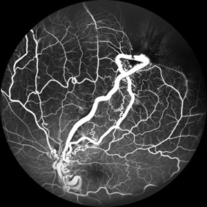 From Artery to Vein, No Detour: Meet the AV Maverick: Racemose Hemangioma
Jul 1 2025 by rohan jain
A case of 10 year old girl with defective vision in LE (6/60) who presented us with this condition.
Photographer: Dr. ROHAN JAIN
Imaging device: mirante
Condition/keywords: arteriovenous malformation, FFA in a case of Racemose angioma, racemose hemangioma
-
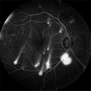 Shooting Stars
Jul 9 2025 by Majda Hadziahmetovic, MD
Fluorescein angiography image demonstrating multiple areas of neovascularization in a middle-aged male patient with long-standing diabetes.
Condition/keywords: proliferative diabetic retinopathy (PDR)
-
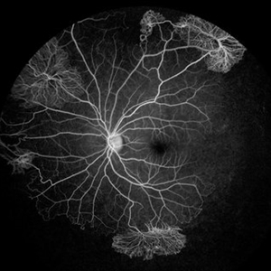 Proliferative Sickle Cell Retinopathy
Jul 8 2025 by Niloofar Piri, MD
Mid AV phase fluorescein angiogram of a 13 yo AA male with SC disease demonstrating multiple classic sea fan neovascularization with peripheral capillary non perfusion (CNP). CNP is more obvious in this image involving the temporal retina and inferonasal retina.
Photographer: Stefan Raev, COT, Saint Louis University
Condition/keywords: Proliferative sickle cell retinopathy, proliferative sickle retinopathy, sickle cell retinopathy
-
 Birdshot Retinochoroidopathy
Jun 18 2025 by César Adrián Gómez Valdivia, MD
Fundus photograph of a 86 YO female patient diagnosed with Birdshot Retinochoroidopathy. Characteristically multifocal cream-colored or yellow-orange, oval or round lesions that emerge from around the optic nerve can be appreciated.
Photographer: @eyemissu2
Imaging device: TOPCON TRC-50DX
Condition/keywords: Birdshot Retinochoroidopathy
-
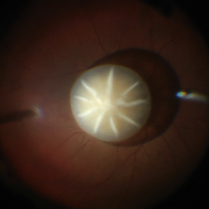 Dislocated Cataractous Lens
Jun 19 2025 by Mrinali Gupta, MD, FASRS
Intraoperative image of a chronically dislocated cataractous lens. The patient underwent pars plana vitrectomy, lensectomy, and placement of an anterior chamber intraocular lens, with improvement in vision from Count Fingers to 20/20 without correction.
Photographer: Mrinali Gupta, MD
Imaging device: Intraoperative surgical video (Zeiss Lumera scope, Resight lens)
Condition/keywords: dislocated crystalline lens
-
 Dislocated Intraocular Lens
Jun 4 2025 by Aditya S Kelkar, MS, FRCS, FASRS,FRCOphth
Fundus photograph of a 79-year-old man with a posteriorly dislocated intraocular lens in the inferior quadrant.
Photographer: Optom Chandrakanta Bhandare, National Institute of Ophthalmology, Pune
Imaging device: Optos Daytona
Condition/keywords: dislocated intraocular lens (IOL)
-
Eye of the Hurricane
Apr 9 2025 by Gustavo Uriel Fonseca Aguirre
Ultrasound biomicroscopy of a post-operative eye (status post trabeculectomy and phacoemulsification) reveals a patent ostium on the right side, along with an intraocular lens in position. A hyphema is observed displaying small convection currents, creating a circular motion pattern due to the temperature gradient between the iris and cornea. Notably, the blood flow can be seen circulating toward the trabeculectomy site.
Condition/keywords: hyphema, trabeculectomy
 Loading…