Discover images
-
 The Peripheral Retina in Profile: A Stereoscopic Atlas
Mar 12 2013 by Norman Byer
The stereoscopic atlas contains unique stereo photographs vividly portraying the changes in the peripheral fundus and their histopathology, incidence and risks.
Condition/keywords: stereo pair, video
-
 Lacquer Cracks
Oct 13 2012 by Geoffrey G. Emerson, MD, PhD, FASRS
Lacquer cracks
Condition/keywords: lacquer cracks, myopic macular degeneration
-
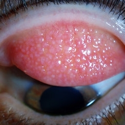
---thumb.JPG/image-square;max$300,300.ImageHandler) Weiss Ring (Floater)
Weiss Ring (Floater)
Jul 10 2013 by Jason S. Calhoun
Patient comes in complaining of a floater towards the nasal aspect of her vision. Fundus photograph with anterior shot, shows a weiss ring pulled off from the optic nerve.
Photographer: Jason S. Calhoun, Department of Ophthalmology, Mayo Clinic Jacksonville, Florida
Condition/keywords: floaters, Weiss ring
-
 Cystic Retinal Tuft
Nov 9 2012 by Norman Byer
This is the same lesion as in the previous slide pair but the photograph was taken nine years later when the patient was 58-years-old soon after an acute posterior vitreous detachment. This demonstrates that posterior vitreous detachment can produce large retinal tears at these sites. However, it is important to emphasize that prophylactic treatment of cystic retinal tufts in the absence of a retinal tear would be very ill-advised because several hundred innocence and harmless lesions would have to be treated in order to prevent one tear of the retina.
Condition/keywords: cystic retinal tuft, posterior vitreous detachment, retinal tear
-
 Giant Papillary Conjunctivitis, Left Upper Eyelid
Jul 22 2013 by Jason S. Calhoun
Contact lens wearer in for exam. Has rough feeling underneath both eyelids. Patient thought it was through SCL wear. Patient VA was 20/20. right eye, 20/30, left eye. Underneath the left upper eyelid, you can see papillary inflammation and redness.
Photographer: Jason S. Calhoun, Department of Ophthalmology, Mayo Clinic Jacksonville, Florida
Imaging device: TOPCON D-90 SL NIKON CAMERA
Condition/keywords: giant papillary conjunctivitis
-
---thumb.jpg/image-square;max$300,300.ImageHandler) CMV Retinitis in a Patient with the Diagnosis of AIDS
CMV Retinitis in a Patient with the Diagnosis of AIDS
Feb 27 2013 by Henry J. Kaplan, MD
CMV retinitis, left eye: classic form in AIDS patient. Hemorrhagic retinitis mainly in the superior arcade.
Condition/keywords: AIDS
-
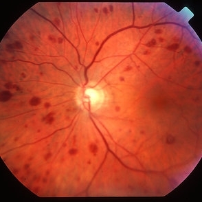
 Severe NPDR
Mar 29 2013 by Henry J. Kaplan, MD
Severe NPDR , IRMA visible inferonasally.
Condition/keywords: nonproliferative diabetic retinopathy
-
 Subhyaloid Hemorrhage
Oct 8 2012 by Jeffrey G. Gross, MD, FASRS
Subhyaloid hemorrhage, layered, with surrounding subretinal hemorrhage.
Condition/keywords: subhyaloid hemorrhage, subretinal hemorrhage
-
 Busacca nodules
May 2 2013 by Henry J. Kaplan, MD
Typical Busacca iris stromal nodules in sarcoid uveitis; notice the ps formation.
Condition/keywords: busacca nodulaes, granulomatous uveitis, iris nodules, sarcoid bussaca iris nodules
-
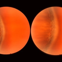 Normal Temporal Ora Serrata
Nov 9 2012 by Norman Byer
This is the normal temporal ora serrata in a 26-year-old man. Note the typical ragged moth-eaten appearance caused by peripheral cystoid degeneration. This appearance may be present in infants but is always present beyond the age of eight years.
Condition/keywords: ora serrata, peripheral cystoid degeneration
-
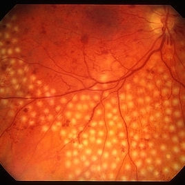
 White Without Pressure
Aug 23 2012 by Gerardo Garcia-Aguirre, MD
Photograph of the temporal peripheral retina showing an area of pale retina (white without pressure).
Photographer: Noemí Hernández, Asociación para Evitar la Ceguera en México
Imaging device: Zeiss FF4
Condition/keywords: pale retina, white without pressure
-
 Siegrist Streaks
Mar 29 2013 by Henry J. Kaplan, MD
Typical Siegrist streaks in hypertensive choridopathy; hyperpigmentations in a linear fashion along choroidal vessels , a rare finding.
Condition/keywords: hypertensive choroidopathy, Siegrist Streaks
-
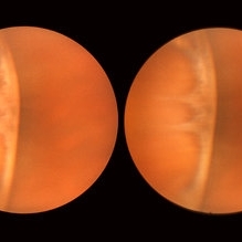 Normal Nasal Ora Serrata
Nov 9 2012 by Norman Byer
This shows the normal nasal ora serrata. Note the dentate processes which divide the nasal ora into prominent bays and teeth
Condition/keywords: dentate processes, normal nasal ora serrata, ora bay, ora teeth
-
 Bilateral Retinoschisis Retinal Detachment
Sep 15 2012 by Barbara Parolini, MD
Fundus photograph of a case of bilateral retinoschisis and retinal detachment. The border of the external layer breaks and the border of the schisis have been treated with argon laser. An epiretinal membrane formed after the formation of retinal detachment.
Photographer: Dr Rino Frisina, Istituto Clinico S.Anna, Brescia, Italy
Imaging device: optos
Condition/keywords: epiretinal membrane formation, retinoschisis
-
 Bergmeister's Papillae
Mar 29 2013 by Henry J. Kaplan, MD
Remnants of fetal hyaloid artery as fibrous tuft called Bergmeister`s papillae on the optic disc.
Condition/keywords: Bergmeister's Papillae, hyaloid artery
-
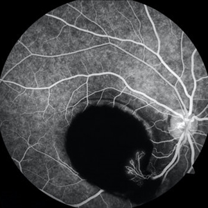 Sub-ILM Hemorrhage with Neovessels
Apr 30 2020 by Saurabh Deshmukh, MBBS, DNB, FVRS, MNAMS
Late arteriovenous phase FA showing a large sub-internal limiting membrane hemorrhage with overlying neovessels. This hypertensive patient presented with a visual acuity of counting fingers at 2 meters. The patient was advised intravitreal anti-VEGF injection, Nd: YAG Membranotomy, and systemic control of hypertension.
Photographer: Saurabh Deshmukh, Sri Sankaradeva Nethralaya, Guwahati, India
Imaging device: Topcon TRC-50 DX
Condition/keywords: hypertensive retinopathy, neovascularization elsewhere (NVE), subILM hemorrhage
-
---thumb.jpg/image-square;max$300,300.ImageHandler) Roth Spot
Roth Spot
Feb 27 2013 by Henry J. Kaplan, MD
Roth spots due to subacute bacterial endocardiris in a patient with the diagnosis of AIDS .
Condition/keywords: AIDS, subacute bacterial endocardiris, white centered retinal hemorrhage (Roth Spot)
-
---thumb.jpg/image-square;max$300,300.ImageHandler) Proliferative Diabetic Retinopathy (PDR) & Traction Retinal Detachment
Proliferative Diabetic Retinopathy (PDR) & Traction Retinal Detachment
Feb 13 2013 by From the Collections of Thomas M. Aaberg, MD and Thomas M. Aaberg Jr., MD
Florid NV with early macular TRD.
Condition/keywords: neovascularization (NV), tractional retinal detachment
-
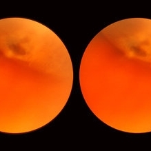
 Open Funnel Retinal Detachment
Oct 13 2012 by Geoffrey G. Emerson, MD, PhD, FASRS
Open funnel retinal detachment
Condition/keywords: B scan ultrasound, open funnel RD
-
 Plaquenil Toxicity
Apr 30 2013 by Theodore Leng, MD, MS, FASRS
SD-OCT scan from a 44-year-old woman with bilateral plaquenil toxicity. There is damage visible in the outer retina in a perifoveal distribution.
Condition/keywords: hydroxychloroquine toxicity, plaquenil toxicity
-
 PRP laser
Mar 29 2013 by Henry J. Kaplan, MD
Right after PRP laser in PDR.
Condition/keywords: laser photocoagulation, pan-retinal photocoagulation (PRP)
-
 Ocular ischaemic syndrome colour 1
Jan 11 2013 by Alex P. Hunyor, MD
Ocular ischaemic syndrome, left eye - color image, posterior pole. Note: dilated but not tortuous veins, attenuated arteries, and multiple intraretinal haemorrhages.
Condition/keywords: ocular ischemic syndrome
-
---thumb.jpg/image-square;max$300,300.ImageHandler) Normal Fundus Photo
Normal Fundus Photo
Feb 13 2013 by From the Collections of Thomas M. Aaberg, MD and Thomas M. Aaberg Jr., MD
Normal fundus photo.
Condition/keywords: fundus photograph, normal eye
-
 Enclosed Ora Bay On The Temporal Side
Nov 9 2012 by Norman Byer
This is another example of an enclosed ora bay on the temporal side. It is surrounded by normal retina and well separated from the ora serrata, which is toward the upper right just beyond the photograph. The yellow nubbin marks an abortive dentate process.
Condition/keywords: abortive dentate process, enclosed ora bay, normal eye, normal retina, ora serrata, temporal retina
-
 Meridional Fold
Nov 9 2012 by Norman Byer
This is the same lesion as in the previous photograph. With the scleral indentation placed more posterior, we now can see that the fold ends over a small collection of subretinal fluid and that there is a very tiny retinal hole just below the posterior end of the retinal fold.
Condition/keywords: peripheral cystoid degeneration, retinal fold, retinal hole, scleral indentation, subretinal fluid
 Loading…