Search results (137 results)
-
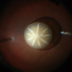 Dislocated Cataractous Lens
Jun 19 2025 by Mrinali Gupta, MD, FASRS
Intraoperative image of a chronically dislocated cataractous lens. The patient underwent pars plana vitrectomy, lensectomy, and placement of an anterior chamber intraocular lens, with improvement in vision from Count Fingers to 20/20 without correction.
Photographer: Mrinali Gupta, MD
Imaging device: Intraoperative surgical video (Zeiss Lumera scope, Resight lens)
Condition/keywords: dislocated crystalline lens
-
Central Retinal Artery Pulsations
May 27 2025 by Malvika Singh
Fundus video showing pulsations of the central retinal artery at the excavated optic nerve head.
Condition/keywords: Central retinal artery, Excavated Disc, Fundus Video
-
Vitreous Waltz vs Retinal Rigidity
Apr 18 2025 by Gustavo Uriel Fonseca Aguirre
B-mode dynamic ultrasound of an eye with vitreous hemorrhage shows hyaloid traction inducing retinal detachment in diabetic retinopathy. The video clearly delineates all anatomical compartments: vitreous, subhyaloid, and subretinal spaces. Characteristic movement patterns are observed - the vitreous demonstrates smooth, wide excursions while the detached retina shows shorter, stiffer motions -confirming tractional pathology.
Condition/keywords: diabetic retinopathy, retinal detachment
-
Endophthalmitis
Apr 9 2025 by Gustavo Uriel Fonseca Aguirre
B-mode ultrasound video of a vitrectomized eye reveals characteristic vitreous cavity membranes secondary to endophthalmitis. The real-time imaging demonstrates these inflammatory membranes exhibit semi-rigid dynamics, displaying viscoelastic behavior with limited displacement during ocular movements while maintaining structural integrity. This restricted mobility pattern, showing both resistance to kinetic forces and slow recoil, represents a pathognomonic feature of advanced fibrotic organization in endophthalmitis.
Condition/keywords: endophthalmitis
-
 Bleb Migration With FAX
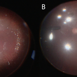
Mar 25 2025 by Robert Andrew Sisk, MD, FACS, FASRS
Color stills from surgical video after subretinal delivery of gene augmentation therapy with voretigene neparvovec-rzyl A) before and B) after fluid-air exchange (FAX). The blebs were between 0.5- and 1-disc diameters from the fovea. After FAX, they gradually extended beneath the fovea and eventually merged. This spared the fovea the trauma from the injection pressure of subretinal injection while allowing treatment to the area.
Imaging device: Leica Proveo 8
Condition/keywords: Fluid-Air Exchange, Gene Therapy, genetic disorder, genetics, Subretinal Injection
-
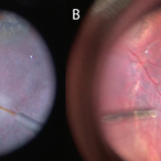
 Completed Bleb with OCT Through Fovea
Mar 25 2025 by Robert Andrew Sisk, MD, FACS, FASRS
Color still from surgical video of subretinal delivery of laru-zova for X-linked retinitis pigmentosa. Live optical coherence tomography (OCT) with foveal tracking via the embedded software in the operating microscope allows monitoring foveal integrity for signs of stress. The contour of the fovea does not exceed the curvature of the bleb (e.g. no inversion). The tangential cannula angle facilitated steering of the bleb posteriorly. The bleb covers essentially the entire macula, which is the target area.
Imaging device: Zeiss Artevo 800
Condition/keywords: gene therapy, genetic disorder, optical coherence tomography (OCT), retinitis pigmentosa, subretinal injection
-
 Cannula Tip Pressure
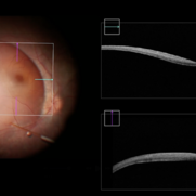
Mar 25 2025 by Robert Andrew Sisk, MD, FACS, FASRS
Color stills from surgical videos of subretinal delivery of gene augmentation therapy with A) voretigene neparvovec-ryzl and B) laru-zova. In the left panel, the cannula is slightly bent, and the retina and RPE are blanched white around the cannula tip engagement. The bleb was challenging to form in this patient with advanced retinal degeneration, and the bleb is shallow and mostly clear. In the right panel, the cannula tip is gently engaged, the cannula is straight, and it follows the retinotomy as the retina is elevated by the injection fluid.
Imaging device: Leica Proveo 8
Condition/keywords: gene therapy, genetic disorder, Leber's congenital amaurosis, retinitis pigmentosa, subretinal injection
-
Macular Hole Surgery: Inverse Flaps
Jan 31 2025 by Thirumalesh Mochi Basavaraj, MD
This video demonstrates, PVD induction , followed by ILM peeling in multiple flower petal flap technique.
Condition/keywords: ILM flaps, ILM peel, induction
-
OCT Video Imaging of Left Eye Age Related Macular Degeneration
Jan 6 2025 by Kavitha Duraipandi, MD DNB FICO FRCS
Left eye OCT macula shows various biomarkers like PED, sub retinal fluid, sub retinal hyper reflective material and hyper reflective foci suggestive of wet age-related macular degeneration.
Condition/keywords: OCT biomarkers, wet age-related macular degeneration (wet AMD)
-
Tractional RD-Making the Decision When and Where to Stop
May 23 2024 by ARVIND JAIN M
This is a young gentlemen with defective vision for 3 months in his right eye. He gave the history of recurrent redness of the right past few months. he was diagnosed to have right eye vasculitis with tractional detachment. He underwent uveitic workup and under steroid cover right eye paraplana vitrectomy with membrane peeling with endolaser with c3f8 gas was planned. patient improved significantly. this surgical video demonstrates when and where to stop during membrane peeling and get good results.
Condition/keywords: Eales disease, retinal vasculitis, tractional retinal detachment
-
Time to Chill
Jan 23 2024 by SHISHIR VERGHESE, MS, FVRS, FAICO (Retina)
Intraoperative surgical video of a 65 year old female patient with advanced proliferative diabetic retinopathy showing neovascularization at the ora serrata for which a cryopexy is being done to cause regression. This video highlights a previously undocumented grape like Neovascularization at the ora serrata in this patient with advanced proliferative diabetic retinopathy.
Condition/keywords: Advanced Proliferative diabetic retinopathy, Cryopexy, neovascularization
-
Retina Rhexis
Jan 2 2024 by Deepak Bhojwani, MS
THIS SURGICAL VIDEO DEMONSTRATES STANDARD ILM PEELING FOR MACULAR HOLE SURGERIES. THE ILM PEELING IS ASSITED BY BRILLIANT BYE DYE STAINING.
Condition/keywords: ILM peeling, macular hole, Macular surgery
-
Smartphone Fundoscopy
Apr 26 2023 by Kalyan Singh
Low cost, portable, high-tech technique of fundoscopy using a smartphone with 20 D or 2.2 panretinal auxiliary lens. This video showing a myopic fundus.
Condition/keywords: fundoscopy, myopia, smartphone
-
Iatrogenic Macular Hole and Subretinal Migration of PFCL
Feb 7 2023 by Aditya S Kelkar, MS, FRCS, FASRS,FRCOphth
The video demonstrates a surgical scenario where the fovea gives away by the force imparted by the jet of an injecting PFCL (Perfluorocarbon heavy Liquid) and the PFCL migrates subfoveally. Intraoperative OCT confirms the presence of a macular hole. The situation is managed by ILM peeling and mobilizing subfoveal PFCL peripherally by injecting another bubble of PFCL over the posterior pole. A peripheral drainage retinotomy is then created to aspirate the subretinal PFCL followed by fluid-air exchange, PFCL-air exchange, and endolaser around the retinotomy. Post-operative OCT at 3 weeks’ follow-up shows a sealed macular hole.
Condition/keywords: Iatrogenic macular hole, Intraoperative complications, Subretinal PFCL
-
Vitrectomy for Macular Hole
Jan 13 2023 by Manish Nagpal, MD, FRCS (UK), FASRS
This is a case of Macular hole for which vitrectomy is being done. After doing core vitrectomy triamcinolone dye is injected to stain the hyaloid. High aspiration is used on cutter to engage the hyaloid and gradually pull it anteriorly. PVD induction is carried out. After this brilliant blue dye is injected to stain the internal limiting membrane. ILM is peeled using a 25 gauge forceps in a tangential manner. After this i use a instrument called the massager which we have developed to gently and atraumatically massage concentrically the edges of the hole. This releases the subtle contaction on the edges of the hole and relaxes the margins. After this air fluid exchange is carried out followed by low vacuum aspiration over the hole. The hole approximates itself gradually as the aspiration dries up the edges.
Condition/keywords: forceps, hyaloid, ILM, macular hole, peeling, staining, video, vitrectomy
-
Vitrectomy for PDR in TRD with Vit haemorrhage
Jan 5 2023 by Manish Nagpal, MD, FRCS (UK), FASRS
Vitrectomy for PDR and TRD and Vitreous haemorrhage using Cutter based dissection. The Vitreous haemorrhage is cleared first. A 25 gauge bevelled cutter is used to dissect all the epiretinal proliferations and tractional components. The ports of these cutters can reach very close to the retinal surface and cut flush without causing any iatrogenic damage to the retinal surface. Forceps is also used to gently peel off a adherent proliferation. Bleeders are stopped raising pressure and applying diathermy. Air fluid exchange is done and viscous subretinal fluid drained from a hole adjacent to diathermy superiorly. Once the retina is flattened endolaser is done 360 degree to achieve long term regression.
Condition/keywords: diabetic retinopathy, diathermy, endolaser, forceps, PDR, peeling, TRD, video, vitrectomy
-
Vitrectomy for PDR for TRD and Subhyaloid Haemorrhage
Jan 4 2023 by Manish Nagpal, MD, FRCS (UK), FASRS
Vitrectomy for PDR and TRD and subhyaloid haemorrhage using Cutter based dissection along with the use of a forceps. The subhyaloid haemorrhage is cleared first using aspiration of the cutter after making a opening in the hyaloid. A 25 gauge bevelled cutter is used to dissect all the epiretinal proliferations and tractional components. The ports of these cutters can reach very close to the retinal surface and cut flush without causing any iatrogenic damage to the retinal surface. Forceps is also used to gently peel off a adherent proliferation. Bleeders are stopped raising pressure and applying diathermy. Once the retina is flattened endolaser is done 360 degree to achieve long term regression.
Condition/keywords: diabetic retinopathy, diathermy, endolaser, forceps, peeling, TRD, video, vitrectomy
-
Vitrectomy for Myopic Retinal Detachment with multiple tears status post prophylaxis
Jan 3 2023 by Manish Nagpal, MD, FRCS (UK), FASRS
Vitrectomy for Myopic Retinal detachment with multiple tears and lattice degenerations status post prophylaxis laser done | Vitrectomy is carried out. Once the vitreous is removed the retina is freely mobile. After this Perfluorocarbon heavy liquid is injected to flatten the posterior pole and push the fluid to the periphery till the edges of the tear. This is followed by endo drainage from infero nasal tear. Scars of laser marks are seen around it. Once the retina flattened endolaser is carried out.
Condition/keywords: air fluid exchange, endo drainage, endolaser, holes, lattice degeneration, myopia, myopic retinal detachment, prophylaxis, RD, reattachment of retinal detachment, tear, video, vitrectomy
-
Vitrectomy for Myopic Retinal Detachment with multiple tears
Jan 2 2023 by Manish Nagpal, MD, FRCS (UK), FASRS
Vitrectomy for Myopic Retinal detachment with multiple tears and lattice degenerations | Vitrectomy is carried out and triamcinolone staining is used to stain the hyaloid attachment. The hyaloid attachment is extremely adherent. With high vacuum the cutter engages the stained hyaloid and gradually peels it off the mobile retina. After this Perfluorocarbon heavy liquid is injected to flatten the posterior pole and push the fluid to the periphery till the edge of the tear. This is followed by endo drainage from the tear. Once the retina flattened endolaser was carried out.
Condition/keywords: air fluid exchange, endo drainage, endolaser, holes, lattice degeneration, myopia, myopic retinal retachment, RD, reattachment of retinal detachment, tear, video, vitrectomy
-
Vitrectomy for Sub ILM blood over macula
Jan 2 2023 by Manish Nagpal, MD, FRCS (UK), FASRS
This is a case of non resolving ILM hemorrhage over macula. Vitrectomy is carried out and hyaloid is removed after traimcinolone staining. After this brilliant blue dye is injected to stain the ILM. Internal limiting membrane is then removed with a forceps. Once the sub ilm blood is exposed , it easily aspirates with the cutter. The origin is probably from a macroaneurysm and there is a small component of subretinal residual blood noted at the end of the surgery.
Condition/keywords: brilliant blue, hyaloid, internal limiting membrane, macula, microaneurysm, retina, sub ILM blood, sub ILM hemorrhage, triamcinolone, video, vitrectomy
-
Vitrectomy for bullous retinal detachment with superior tear
Jan 2 2023 by Manish Nagpal, MD, FRCS (UK), FASRS
Vitrectomy for bullous Retinal detachment with superior tear| In this case vitrectomy is being done for a retinal detachment with superior tear. Once the vitreous is removed, air fluid exchange is carried out. Perfluorocarbon heavy liquid is injected to flatten the posterior pole and push the fluid to the periphery for endo drainage. This is followed by endo drainage from the superior break. Once the retina flattened endolaser was carried out.
Condition/keywords: air fluid exchange, bullous retinal detachment, endo drainage, endolaser, holes, Prophylaxis, RD, reattachment of retinal detachment, tear, video, vitrectomy
-
Vitrectomy for Sub ILM blood over macula
Jan 2 2023 by Manish Nagpal, MD, FRCS (UK), FASRS
This is a case of non resolving ILM haemorrhage over macula. Vitrectomy is carried out and hyaloid is removed. Cutter is used to try to aspirate the blood from the surface of macula. But due to its location in the sub ILM space i use a forceps to make a opening in the ILM. Through the opening the blood aspirates easily.
Condition/keywords: hyaloid, internal limiting membrane, macula, retina, sub ILM blood, sub ILM hemorrhage, video, vitrectomy
-
Vitrectomy for retinal detachment with superior holes
Jan 2 2023 by Manish Nagpal, MD, FRCS (UK), FASRS
Vitrectomy for Retinal detachment with multiple holes superiorly | In this case vitrectomy is being done for a retinal detachment with superior holes. Once the vitreous is removed, air fluid exchange is carried out. This is followed by endo drainage from the superior break. Once the retina flattened endolaser was carried out.
Condition/keywords: air fluid exchange, bullous retinal detachment, endo drainage, endolaser, holes, Prophylaxis, RD, reattachment of retinal detachment, video, vitrectomy
-
Vitrectomy TRD in Proliferative diabetic retinopathy
Jan 2 2023 by Manish Nagpal, MD, FRCS (UK), FASRS
Vitrectomy for PDR and TRD using Cutter based dissection| This is a case of subhyaloid hemorrhage and Tractional retinal detachment in a diabetic patient. The subhyaloid hemorrhage is aspirated using the cutter . 25 gauge bevelled cutter is used to dissect all the epiretinal proliferations and tractional components. The ports of these cutters can reach very close to the retinal surface and cut flush without causing any iatrogenic damage to the retinal surface. Forceps are used to peel adherent membranes Bleeders are stopped raising pressure and applying diathermy. Once the retina is flattened endolaser is done 360 degree to achieve long term regression.
Condition/keywords: cutter, diabetic retinopathy, endolaser, forceps, PDR, peeling, proliferative diabetic retinopathy (PDR), PRP, tractional retinal detachment, TRD, video, vitrectomy
-
Vitrectomy for PDR in TRD
Dec 16 2022 by Manish Nagpal, MD, FRCS (UK), FASRS
This is a case of subhyaloid haemorrhage and Tractional retinal detachment in a diabetic patient. The subhyaloid haemorrhage is aspirated using the cutter . 25 gauge bevelled cutter is used to dissect all the epiretinal proliferations and tractional components. The ports of these cutters can reach very close to the retinal surface and cut flush without causing any iatrogenic damage to the retinal surface. Bleeders are stopped raising pressure and applying diathermy. Once the retina is flattened endolaser is done 360 degree to achieve long term regression.
Condition/keywords: cutter, proliferative diabetic retinopathy (PDR), video, vitrectomy
 Loading…