Search results (137 results)
-
 The Peripheral Retina in Profile: A Stereoscopic Atlas
Mar 12 2013 by Norman Byer
The stereoscopic atlas contains unique stereo photographs vividly portraying the changes in the peripheral fundus and their histopathology, incidence and risks.
Condition/keywords: stereo pair, video
-
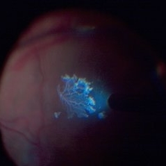 Detached NVE During PVD induction
Apr 27 2018 by Michael J. Koss, MD, PhD, MBA
A 73-year-old woman with macular pucker underwent a pars plana vitrectomy with membrane peeling. Additionally the patient suffers from diabetic retinopathy after being diagnosed with type 2 diabetes mellitus sixteen years ago. Prior to the procedure she was treated with a series of intravitreal Bevacizumab-injections due to diabetic macular edema. There was no history of a proliferative DRP. During the vitrectomy a branch of an obliterated NVE spontaneously detached and floated freely in the vitreous. The 3D shot was captured via Alcon’s NGENUITY® 3D Visualization System in form of photograph and video providing an outstandingly detailed image of the branched NVE.
Photographer: Michael Koss, Augenzentrum Nymphenburger Hoefe
Imaging device: Alcon’s NGENUITY® 3D Visualization System
Condition/keywords: diabetes, diabetic retinopathy, neovascularization elsewhere (NVE), pars plana vitrectomy (PPV), PVD induction
-
Epiretinal membrane removal
Oct 24 2022 by Manish Nagpal, MD, FRCS (UK), FASRS
This video highlights the surgical technique of tangentially removing the epiretinal membrane using a forceps
Photographer: Manish Nagpal
Condition/keywords: epiretinal membrane, ERM, macular pucker, staining, video, vitrectomy
-
Flattening a bullous retinal detachment
Oct 24 2022 by Manish Nagpal, MD, FRCS (UK), FASRS
This surgical clip shows the way a bullous retinal detachment reattaches at the time of endodrainage
Photographer: Manish Nagpal
Condition/keywords: bullous detachment, endodrainage, tear, video, vitrectomy
-
Flattening of Giant retinal tear
Oct 24 2022 by Manish Nagpal, MD, FRCS (UK), FASRS
This video highlights the step of eversion of the giant retinal tear flap and flattening of retina using heavy PFCL liquid
Photographer: Manish Nagpal
Condition/keywords: flap, giant retinal tear, GRT, PFCL, video, vitrectomy
-
Iatrogenic Macular Hole and Subretinal Migration of PFCL
Feb 7 2023 by Aditya S Kelkar, MS, FRCS, FASRS,FRCOphth
The video demonstrates a surgical scenario where the fovea gives away by the force imparted by the jet of an injecting PFCL (Perfluorocarbon heavy Liquid) and the PFCL migrates subfoveally. Intraoperative OCT confirms the presence of a macular hole. The situation is managed by ILM peeling and mobilizing subfoveal PFCL peripherally by injecting another bubble of PFCL over the posterior pole. A peripheral drainage retinotomy is then created to aspirate the subretinal PFCL followed by fluid-air exchange, PFCL-air exchange, and endolaser around the retinotomy. Post-operative OCT at 3 weeks’ follow-up shows a sealed macular hole.
Condition/keywords: Iatrogenic macular hole, Intraoperative complications, Subretinal PFCL
-
 OCT Artifacts
Dec 5 2012 by Yale L. Fisher, MD
Dr. Jay Duker examines the critical issue of OCT artifacts and discusses how to identify and remedy them. NOTE: This movie is based on a live lecture and contains a few minor audio defects- they're not significant enough to interfere with your viewing experience and should not be confused with any problems with your viewing system. Dr. Duker's Financial Interest Disclosure: Stockholder- Hemera Biosciences Research Support- OptoVue Carl Zeiss Meditech Topcon Scientific Advisory Board- Paloma Pharmaceuticals Consultant- Alcon Genentech Ophthotech Novartis Neovista
Condition/keywords: video
-
 Pars Plana Cysts
Jul 29 2020 by Vinod Kumar
Pars plana cysts found during peripheral indentation at the conclusion of vitrectomy for rhegmatogenous retinal detachment.
Photographer: Vinod Kumar
Imaging device: Still from a video
Condition/keywords: cyst of the pars plana
-
 Remnant of Hyaloidal Artery
Feb 5 2014 by Gerardo Garcia-Aguirre, MD
Video of the fundus of the left eye of a 14-year-old asymptomatic female, where a prepapillary vitreous opacity is observed. The opacity is attached to the origin of the retinal vessels in the optic nerve head, and is considered to be a remnant of the hyaloidal artery.
Photographer: Gerardo Garcia-Aguirre, MD
Condition/keywords: persistence of the hyaloid artery
-
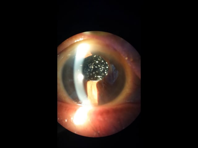
Spontaneous lens dislocation (Weill Marchesani Syndrome)
Nov 9 2022 by Heitor Nogueira
A 9 year-old Male patient diagnosed with Weill Marchesani presented spontaneous bilateral lens dislocation. Weill-Marchesani syndrome, also known as spherophakia-brachymorphy syndrome and mesodermal dysmorphodystrophy, is an inherited connective tissue disorder characterized by eye lens abnormalities, secondary glaucoma, short stature, brachydactyly, joint stiffness, and cardiovascular defects.
Photographer: Heitor Nogueira, Insituto Penido Burnier, Campinas-SP, Brazil
Condition/keywords: mesodermal dysmorphodystrophy, spherophakia-brachymorphy syndrome, spontaneous lens dislocation, video, Weill Marchesani Syndrome
-
Time to Chill
Jan 23 2024 by SHISHIR VERGHESE, MS, FVRS, FAICO (Retina)
Intraoperative surgical video of a 65 year old female patient with advanced proliferative diabetic retinopathy showing neovascularization at the ora serrata for which a cryopexy is being done to cause regression. This video highlights a previously undocumented grape like Neovascularization at the ora serrata in this patient with advanced proliferative diabetic retinopathy.
Condition/keywords: Advanced Proliferative diabetic retinopathy, Cryopexy, neovascularization
-
Vitrectomy for bullous retinal detachment with superior tear
Jan 2 2023 by Manish Nagpal, MD, FRCS (UK), FASRS
Vitrectomy for bullous Retinal detachment with superior tear| In this case vitrectomy is being done for a retinal detachment with superior tear. Once the vitreous is removed, air fluid exchange is carried out. Perfluorocarbon heavy liquid is injected to flatten the posterior pole and push the fluid to the periphery for endo drainage. This is followed by endo drainage from the superior break. Once the retina flattened endolaser was carried out.
Condition/keywords: air fluid exchange, bullous retinal detachment, endo drainage, endolaser, holes, Prophylaxis, RD, reattachment of retinal detachment, tear, video, vitrectomy
-
Vitrectomy for PDR in TRD
Dec 16 2022 by Manish Nagpal, MD, FRCS (UK), FASRS
This is a case of subhyaloid haemorrhage and Tractional retinal detachment in a diabetic patient. The subhyaloid haemorrhage is aspirated using the cutter . 25 gauge bevelled cutter is used to dissect all the epiretinal proliferations and tractional components. The ports of these cutters can reach very close to the retinal surface and cut flush without causing any iatrogenic damage to the retinal surface. Bleeders are stopped raising pressure and applying diathermy. Once the retina is flattened endolaser is done 360 degree to achieve long term regression.
Condition/keywords: cutter, proliferative diabetic retinopathy (PDR), video, vitrectomy
-
 A Motor Vehicle Accident Causing Valsalva Retinopathy OD, While Racing A Side By Side 4 Wheel Off-Road Vehicle
Apr 29 2020 by John S. King, MD
43-year-old white male who was injured while racing a side by side 4-wheel off-road vehicle (see Video: https://imagebank.asrs.org/file/53854/sxs-crash-during-a-race-causing-valsalva-retinopathy-od). He presented about three weeks after the injury. He was being seen by his local eye doctor who wanted an evaluation for the retinal heme and scotoma. His main complaint was a central/parcentral scotoma described as a greyish area in vision. Va 20/50 OD, nomotensive, no APD (by technician), anterior segment u/r; see picture for the fundus exam - of note there are superficial/preretinal heme, with layering of the heme superiorly, and small superficial heme at nasal edge of the optic disc; in the parafoveal region nasally there is some mottling of the RPE that may indicate an area of prior commotio retinae (also possible to have TON), which may account for his scotoma. Really bad accident (video), and amazingly, he had no LOC or injuries other than the right retina. Helmet and racing harness seat belt were used.
Photographer: Asli Ahmed
Imaging device: Topcon 50
Condition/keywords: valsalva retinopathy
-
 A Motor Vehicle Accident Causing Valsalva Retinopathy OD, While Racing A Side By Side 4 Wheel Off-Road Vehicle
May 5 2020 by John S. King, MD
A 43-year-old white male who was injured while racing his side by side 4 wheel off-road vehicle (this is a video he showed me on his phone). He presented about three weeks after the injury. He was being seen by his local eye doctor who wanted an evaluation for the retinal heme and scotoma. His main complaint was a central/parcentral scotoma described as a greyish area in vision. Va 20/50 OD, nomotensive, no APD (by technician), anterior segment u/r; see {https://imagebank.asrs.org/file/53828/sxs-crash-during-a-race-causing-valsalva-retinopathy-od} for the fundus exam - of note there are superficial/preretinal heme, with layering of the heme superiorly; in the parafoveal region nasally there is some mottling of the RPE that may indicate an area of prior commotio retinae (also possible to have TON), which may account for his scotoma. Really bad accident, and amazingly, he had no LOC or injuries other than the right retina. Helmet and racing harness seat belt were used.
Condition/keywords: motor vehicle accident, trauma, valsalva retinopathy
-
 Asteroid Hyalosis, Posterior Vitreous Separation
Dec 10 2012 by Yale L. Fisher, MD
VA 20/20. In asteroid hyalosis, there is a space between the posterior hyaloid face and the calcified asteriod lesions. Use gain to differentiate between VH and asteroid hyalosis (asteroid still present when gain is decreased).
Condition/keywords: video
-
 25G PPV Without Scleral Buckling for RRD, PVR, Giant Breaks
Dec 10 2012 by Yale L. Fisher, MD
Dr. Steve Charles shares his approach to 25G PPV without scleral buckling for RRD, PVR and giant breaks. NOTE: A narration by Dr. Steve Charles will soon be available for this movie- please check back periodically.
Condition/keywords: video
-
 25G Vitrectomy for Diabetic Tractional Retinal Detachment
Dec 10 2012 by Yale L. Fisher, MD
Dr. Steve Charles discusses his successful approach to performing vitrectomy for diabetic tractional retinal detachment using 25 gauge.
Condition/keywords: video
-
 25G Vitreomacular Surgery
Dec 10 2012 by Yale L. Fisher, MD
Pearls of wisdom from Dr. Steve Charles for surgeons examining their approach to 25G vitreomacular surgery. NOTE: A narration by Dr. Steve Charles will soon be available for this movie- please check back periodically.
Condition/keywords: video, vitreomacular surgery
-
 27-Gauge Vitrectomy
Aug 3 2013 by Yusuke Oshima, MD, PhD
A high-performance 27-gauge vitrectomy system (27+) was used for treating a case with primary rhegmatogenous retinal detachment complicated with macular hole.
Condition/keywords: macular hole, video, vitrectomy
-
 Acute Partial Vitreous Separation
Dec 10 2012 by Yale L. Fisher, MD
This is a partial vitreous separation that demonstrates a mildly reflective curvilinear shape of the partially separated vitreous face. The vitreous is attached inferonasally and inferotemporally, but detached and freely mobile at 6 o'clock.
Condition/keywords: video
-
 Asteroid hyalosis
Aug 26 2015 by René Hernán Parada Vásquez
It is a video of a 60-year-old male with an asteroid hyalosis, this is a multiple-yellow-white, round, particles composed of calcium. You can see the movement of the vitreous when the patient looks up and down.
Photographer: Parada René, ESO, Guatemala
Condition/keywords: asteroid hyalosis, floaters
-
 Asteroid Hyalosis, Vitreous Face Attached
Dec 10 2012 by Yale L. Fisher, MD
In asteroid hyalosis, accumulations of calcium soaps dispersed throughout the vitreous produce bright echoes in the usually echolucent vitreous. The appearance of asteroid hyalosis should not be confused with that of vitreous hemorrhage or vitritis. Many of the larger aggregates in asteroid hyalosis are easily seen as the gain is reduced to below 60 db, unlike vitreous hemorrhage or vitritis which usually disappears at low gain settings. There is also an area of clear echolucent vitreous between the posterior hyaloid face and the asteroid particles, which is usually not present in vitreous hemorrhage or vitritis.
Condition/keywords: video
-
 Attached Vitreous With Floaters
Dec 10 2012 by Yale L. Fisher, MD
The vitreous is attached and demonstrates after-movements of formed vitreous as the patient is asked to look to the right and left. There is mild reflectivity in the formed vitreous from collagen. The optic nerve is visible in the superior aspect of the image and the lateral rectus muscle is seen inferiorly.
Condition/keywords: floaters, video, vitreous
-
Bimanual Tractional Retinal Detachment Repair with Viscous Subretinal Fluid Removal
Nov 28 2022 by Nimesh A. Patel, MD, FASRS
Using a chandelier endoillumination, a 2-handed approach with forceps and a vitrector can be used to remove preretinal membranes in a TRD from PDR. The subretinal fluid had a high viscosity. It was removed mechanically by rotating the vitrector.
Condition/keywords: subretinal fluid, video
 Loading…