Search results (146 results)
-
 Necrotizing Scleritis
Apr 17 2025 by Gustavo Uriel Fonseca Aguirre
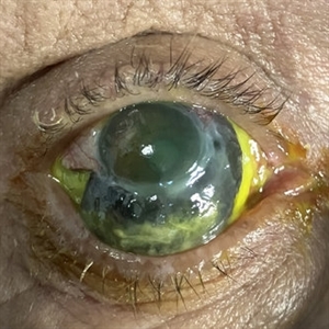
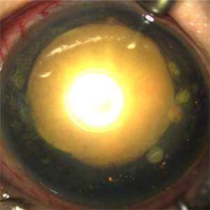
The clinical photograph shows necrotizing scleritis with perilimbal involvement, featuring marked scleral thinning and violaceous episcleral injection in the inferior quadrant. Focal uveal prolapse is visible at the area of maximal scleral necrosis, accompanied by peripheral ulcerative keratitis. Fluorescein staining residue is observed on the ocular surface. Associated findings include mild conjunctival chemosis and dilated episcleral vessels.
Photographer: Gustavo U. Fonseca Aguirre, Hospital Conde de Valenciana, Ciudad de México
Condition/keywords: necrotizing scleritis
-
 Cheese Pizza Pie Appearance in CMV Retinitis
Mar 30 2024 by KANWALJEET HARJOT MADAN, M.S. (Ophthalmology), FAICO (Vitreous - Retina)
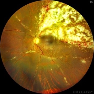
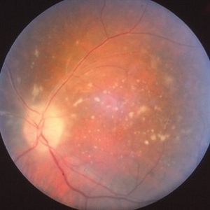
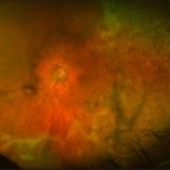
This is Fundus Photograph of left eye of 53 year male depicting an area of Retinal Necrosis with few Retinal Haemorrhages suggestive of CMV Retinitis. Areas of Perivascular Exudation also seen. On investigations, the patient was found to be HIV positive. He was started on Anti Retro Viral treatment after physician opinion.
Photographer: Dr. Kanwaljeet Harjot Madan, Thind Eye Hospital, Jalandhar City (Punjab) INDIA.
Imaging device: Zeiss Fundus Camera
Condition/keywords: AIDS, cytomegalovirus (CMV), retinitis
-
 Pigmented KPs
Dec 8 2023 by Nassim Alejandro Abreu Arbaje, MD
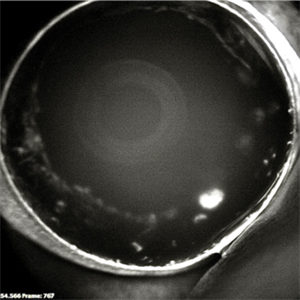
Anterior segment photograph of a 27 year old female diagnosed with an Acute Retinal Necrosis. In the picture we can see mutton fat keratic precipitates already pigmented.
Photographer: Nassim Abreu
Imaging device: Alcon NGenuity Systems
Condition/keywords: Acute Retinal Necrosis, mutton-fat keratic precipitates (KP), Uveitis
-
 CMV retintis
Sep 14 2023 by Ben Serar
Fundus photograph of LE showing superficial flame-shaped haemorrhages with surrounding retinal necrosis at the posterior pole along the superotemporal arcade in a case of fulminant type of CMV retinitis.
Condition/keywords: CMV retinitis, fulminant retinitis, pizza-pie appearance, viral retinitis
-
 Acute Retinal Necrosis with Retinal Detachment
Jan 13 2022 by Tuan Tran, MBBS, MMed (OphthSc), FRANZCO, DRCPSC
Acute Retinal Necrosis with Retinal Detachment.
Photographer: Tuan Tran
Condition/keywords: Acute Retinal Necrosis with Retinal Detachment, retinal necrosis
-
 Left Acute Retinal Necrosis
Jan 11 2022 by Tuan Tran, MBBS, MMed (OphthSc), FRANZCO, DRCPSC
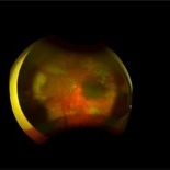
84 year-old gentleman presenting with left acute retinal necrosis.
Photographer: Tuan Tran
Imaging device: Optos widefield
Condition/keywords: ARN complications
-
 Necrotic Multifocal Retinoblastoma Group E (ICRB) / cT3e (AJCC)
Jul 7 2021 by Linda A Cernichiaro- Espinosa, MD
A 3-year, 9-month-old male presented with unilateral advanced group E multifocal retinoblastoma cT3e (AJCC). Anterior seeding vascularized over the iris surface. Fluorescein angiogram fills the vascularized tumors. Aseptic orbital cellulitis, birefringent anterior segment crystals, cataract and dense vitritis are secondary to necrosis.
Photographer: Jose Oyervides-Alvarado MD
Imaging device: RetCam3
Condition/keywords: fluorescein angiogram (FA), pediatric tumor, retinoblastoma
-
 Necrotic Multifocal Retinoblastoma Group E (ICRB) / cT3e (AJCC)
Jul 7 2021 by Linda A Cernichiaro- Espinosa, MD
A 3-year, 9-month-old male presented with unilateral advanced group E multifocal retinoblastoma cT3e (AJCC). Anterior seeding vascularized over the iris surface. Fluorescein angiogram fills the vascularized tumors. Aseptic orbital cellulitis, birefringent anterior segment crystals, cataract and dense vitritis are secondary to necrosis.
Photographer: Jose Oyervides-Alvarado MD
Imaging device: RetCam3
Condition/keywords: retinoblastoma
-
 Acute Retinal Necrosis
May 31 2021 by Aditya S Kelkar, MS, FRCS, FASRS,FRCOphth
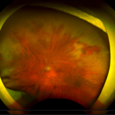
Fundus photograph of 43-year-old female with left eye acute retinal necrosis.
Imaging device: Clarus 500
Condition/keywords: acute retinal necrosis
-
 Optic Disc Melanocytoma
Mar 15 2021 by Deepak Bhojwani, MS
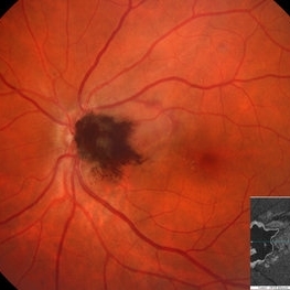
Fundus photograph of a 49- year-old gentlemen with a characteristic dark brown elevated pigmented mass lesion centered on optic disc and extending into temporal peripapillary area classically suggestive of optic disc melanocytoma. Also note the pigment dispersion and retinal edema just superotemporal to the lesion secondary to tumor necrosis. Inset -Enface OCT image segmented at IS-OS ellipsoid zone level delineating exact horizontal & vertical extent of this tumor mass. Enface OCT imaging also helps in detailing the choroidal extension of such tumors.
Photographer: DEEPAK BHOJWANI; OCCURA EYE CARE & RESEARCH CENTER
Imaging device: OCT
Condition/keywords: enface imaging, melanocytoma, optic disc, optical coherence tomography (OCT)
-
 Blunt Ocular Trauma Due to Firework Injury
Jun 9 2020 by Brittany Rota
Ultra- widefield pseudocolor image of an 18-year-old male with blunt ocular trauma in the right eye due to a firework injury. The patient presented with commotio retinae (sclopteria), an acute vitreous hemorrhage, choroidal rupture, and a subretinal hemorrhage. The referring physician performed surgery on the lateral rectus muscle which was macerated but not severed, and several orbital fibrous foreign bodies were removed from the posterior orbit. The globe was intact. There is no evidence of retinal tear in the region of sclopetaria; however, there is complete necrosis of the temporal peripheral choroid and retina. The vitreous hemorrhage was slowly clearing on his exam 6-9-2020. The patient is developing subretinal fibrosis. The physician is concerned about the choroidal rupture that is visible through the submacular hemorrhage. There is one rupture that appears to course directly under the fovea. The physician states that if this is the case, his vision most likely will be 20/200 or worse. His vision was hand motion in all fields except nasally, which he was unable to see hand motion at his visit on 6-9-2020.
Photographer: Brittany Rota
Imaging device: Optos California
Condition/keywords: blunt trauma, choroidal rupture, commotio retinae, fibrosis, firework injury, fundus photograph, hand motion, necrotizing retina, Optos, pseudocolor, subretinal hemorrhage, vitreous hemorrhage
-
 Mushroom-shaped Choroidal Melanoma
May 18 2020 by McGill University Health Centre
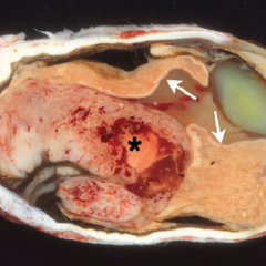
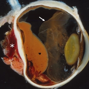
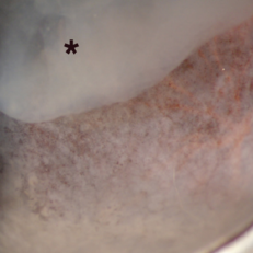
The enucleation specimen in (A) shows a diffuse melanoma infiltrating the choroid and ciliary body. In the center, a large area of necrosis and hemorrhage is present (*) and the retina is infiltrated (arrows). Note hypopyon in the anterior chamber and the cataractous lens.
Condition/keywords: enucleation, mushroom-shaped
-
 Amelanotic Choroidal Melanoma
May 18 2020 by McGill University Health Centre
The enucleation image shows a large amelanotic tumor with large areas of hemorrhage and necrosis. Note the several dilated blood vessels and an adjacent retinal detachment with lipofuscin pigment on its surface (arrow).
Condition/keywords: amelanotic melanoma, enucleation, mushroom-shaped
-
 Choroidal Melanoma with Extraocular Extension
May 18 2020 by McGill University Health Centre
Choroidal melanoma is often asymptomatic and diagnosis is incidental. The tumors may grow beneath the retina, or may break through the Bruch membrane and disrupt the retina. Tumors breaking through the Bruch membrane and disrupting the retina have a characteristic “mushroom” shape. The diffuse melanoma seen in this enucleation specimen is infiltrating the choroid. Extensive areas of necrosis and hemorrhage are present (arrowhead). The extraocular involvement is large and surrounded by hemorrhage (*). The retina is folded (arrow).
Condition/keywords: extraocular extension
-
 Two-Lobed Melanotic and Amelanotic Tumor
May 18 2020 by McGill University Health Centre
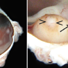
Choroidal melanoma is often asymptomatic and diagnosis is incidental. The tumors may grow beneath the retina, or may break through the Bruch membrane and disrupt the retina. Tumors breaking through the Bruch membrane and disrupting the retina have a characteristic “mushroom” shape. The enucleation specimen in (A) shows a 2-lobed melanotic (arrow) and amelanotic (arrowhead) tumor in the posterior pole of the eye overlying the optic nerve head. In (B), higher magnification shows the optic nerve head and a feeder tumor vessel (arrow). Necrosis is present in the amelanotic tumor (arrowheads).
Condition/keywords: enucleation, tumor
-
 Ciliary Body Melanoma
May 18 2020 by McGill University Health Centre
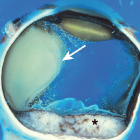
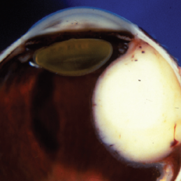
Uveal melanoma is the most common primary eye malignancy in adulthood, occurring mainly after age 60. The uveal tract — composed of the iris, ciliary body, and choroid — can be affected by uveal melanoma. Despite advances in treatment of the primary tumor, metastatic disease occurs in almost half of patients, generally affecting the liver and lungs via hematogenous dissemination of the primary tumor. Tumors have different levels of pigmentation, and some are amelanocytic (nonpigmented). The differential diagnosis for amelanotic choroidal melanoma is metastatic disease. Large tumors displace the lens. Of the 3 locations in the uveal tract, tumors of the ciliary body have the worst prognosis. This enucleation specimen shows a pigmented, nodular-shaped ciliary body melanoma (arrow) with extensive necrosis (*). A retinal detachment is present with subretinal fluid (arrowhead), and the retina is folded (•).
Condition/keywords: enucleation, melanoma
-
 Melanocytoma of the Ciliary Body and Choroid
May 18 2020 by McGill University Health Centre
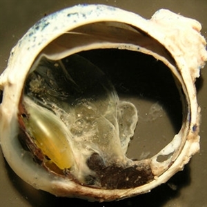
This type of melanocytoma, which tends to undergo spontaneous necrosis, is a variant of melanocytic nevi of the uveal tract. Follow-up is recommended due to the possibility of malignant transformation (melanoma), which occurs in up to 2% of cases. Gross examination reveals a very pigmented, solid, homogeneous tumor located in the ciliary body and choroid. Tumor thickness and pigmentation suggests uveal melanoma on gross examination; however, the heavy pigmentation can indicate melanocytoma. Diagnosis is confirmed by microscopic examination. This image shows a transverse section of an enucleation specimen. A thick melanocytic tumor is present in the uveal tract, at the retina and near the ciliary bodies
Condition/keywords: choroid, enucleation, melanocytoma
-
 Metastatic Adenocarcinoma
May 18 2020 by McGill University Health Centre
Metastatic disease is the most frequent intraocular malignant tumor. In women, the most common origin is breast cancer. In men, the most common origin is lung cancer. This pupil–optic nerve section shows a whitish tumor with several foci of necrosis (*) occupying the posterior aspect of the choroid. Note the pigment epithelium over the inner surface of the tumor. A serous retinal detachment is present (arrow) with a retinal detachment artifact overlying the tumor and normal choroid. Note the air bubble artifacts in the vitreous cavity. Another artifact, the compression of the eyeball, is present on the right side.
Condition/keywords: breast cancer, foci of necrosis, metastatic adenocarcinoma, tumor
-
 Leiomyoma
May 18 2020 by McGill University Health Centre
Leiomyoma is a benign, smooth muscle tumor. Ninety percent of cases occur in women. The differential diagnosis includes amelanotic melanoma and nerve sheath tumors. This transversal pupil–optic nerve (PO) section of an enucleation specimen shows a nodular, well-delineated, whitish tumor in the ciliary body. The cut surface shows small foci of hemorrhage without necrosis. The retina partially covers the inner surface of the tumor, and the sclera is not infiltrated. Note the slightly displaced (subluxated) cataractous lens and the choroidal detachment artifact in the right inferior corner.
Condition/keywords: enucleation, leiomyoma, tumor
-
 Toxoplasmic Acute Retinal Necrosis
May 18 2020 by McGill University Health Centre
Toxoplasmosis can have several manifestations in the eye, of which toxoplasmic acute retinal necrosis has the worst prognosis. This enucleation specimen shows extensive retinal necrosis with multiple coalescent foci. The vitreous is hazy (*).
Condition/keywords: acute retinal necrosis, toxoplasmosis
-
 Large B Cell Lymphoma of the Retina
Dec 13 2019 by McGill University Health Centre
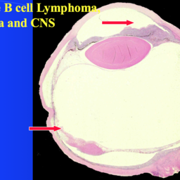
65-year-old female with the clinical diagnosis of bilateral uveitis of unknown etiology. Histopathology of the enucleated specimen showing large neoplastic cells in the anterior chamber, representing pseudohypopyon in a case of masquerade syndrome. The retina shows areas of necrosis with neoplastic large B cells.
Photographer: Miguel N. Burnier, McGill University Health Center-McGill University Ocular Pathology & Translational Research Laboratory
Imaging device: Zeiss
Condition/keywords: large b cell lymphoma, large b cell lymphoma of the retina, masquerade syndrome, pesudohypopyon
-
 CMV Retinitis in AIDS Patient
Dec 12 2019 by McGill University Health Centre
Fundus photograph of a 32-year-old man with HIV infection and 100 CD4+ cells count. Several areas of retinal necrosis interspersed with areas of hemorrhage around blood vessels can be observed.
Photographer: Miguel N. Burnier, McGill University Health Center-McGill University Ocular Pathology & Translational Research Laboratory
Imaging device: Fundoscopy
Condition/keywords: AIDS, cytomegalovirus (CMV), HIV, retinitis
-
 Primary Intraocular Lymphoma
Nov 20 2019 by McGill University Health Centre
73-year-old man with retinal vasculitis and acute retinal lesions of the left eye. Optic nerve and retinal infiltrates consistent with acute retinal necrosis.
Condition/keywords: acute retinal necrosis, primary intraocular lymphoma
-
 Progressive Outer Retinal Necrosis
Nov 5 2019 by Nichole Lewis
86-year-old male with progressive outer retinal necrosis, significant retinitis, retinal whitening, intraretinal hemorrhages and peripheral rpe changes. FA showed occlusive vasculitis with non-perfusion. Patient is immuno-suppressed with a history of renal transplant. VA 20/60.
Photographer: Nichole Lewis
Imaging device: Optos
Condition/keywords: intraretinal hemorrhage, occlusive vasculitis, progressive outer retinal necrosis (PORN), retinal pigment epithelium (RPE) changes, retinal whitening, retinitis
 Loading…