 Initializing download.
-
 By McGill University Health Centre
The MUHC-McGill University
Co-author(s): Sabrina Bergeron, P. Zoroquiain, E. Esposito, S. Corredor Casas, P. Logan, A. N. Odashiro, Miguel N. Burnier, Paulina García de Alba Graue, McGill University Health Center-McGill University Ocular Pathology & Translational Research Laboratory - Uploaded on May 18, 2020.
- Last modified by Caroline Bozell on May 19, 2020.
- Rating
- Appears in
- Ciliary Body Melanoma
- Condition/keywords
- melanoma, enucleation
- Description
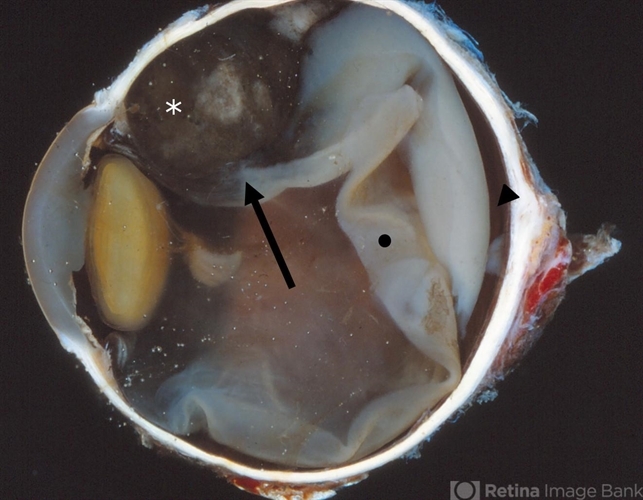
- Uveal melanoma is the most common primary eye malignancy in adulthood, occurring mainly after age 60. The uveal tract — composed of the iris, ciliary body, and choroid — can be affected by uveal melanoma. Despite advances in treatment of the primary tumor, metastatic disease occurs in almost half of patients, generally affecting the liver and lungs via hematogenous dissemination of the primary tumor. Tumors have different levels of pigmentation, and some are amelanocytic (nonpigmented). The differential diagnosis for amelanotic choroidal melanoma is metastatic disease. Large tumors displace the lens. Of the 3 locations in the uveal tract, tumors of the ciliary body have the worst prognosis. This enucleation specimen shows a pigmented, nodular-shaped ciliary body melanoma (arrow) with extensive necrosis (*). A retinal detachment is present with subretinal fluid (arrowhead), and the retina is folded (•).


