 Initializing download.
-
 By McGill University Health Centre
The MUHC-McGill University
Co-author(s): Sabrina Bergeron, P. Zoroquiain, E. Esposito, S. Corredor Casas, P. Logan, A. N. Odashiro, Miguel N. Burnier, Paulina García de Alba Graue, McGill University Health Center-McGill University Ocular Pathology & Translational Research Laboratory - Uploaded on May 18, 2020.
- Last modified by Caroline Bozell on May 19, 2020.
- Rating
- Appears in
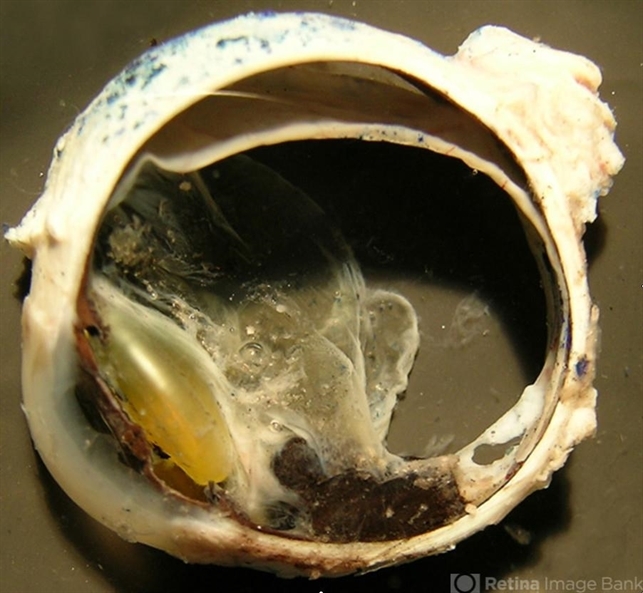
- Melanocytoma
- Condition/keywords
- melanocytoma, choroid, enucleation
- Description
- This type of melanocytoma, which tends to undergo spontaneous necrosis, is a variant of melanocytic nevi of the uveal tract. Follow-up is recommended due to the possibility of malignant transformation (melanoma), which occurs in up to 2% of cases. Gross examination reveals a very pigmented, solid, homogeneous tumor located in the ciliary body and choroid. Tumor thickness and pigmentation suggests uveal melanoma on gross examination; however, the heavy pigmentation can indicate melanocytoma. Diagnosis is confirmed by microscopic examination. This image shows a transverse section of an enucleation specimen. A thick melanocytic tumor is present in the uveal tract, at the retina and near the ciliary bodies


