 Initializing download.
-
 By McGill University Health Centre
The MUHC-McGill University
Co-author(s): Sabrina Bergeron, P. Zoroquiain, E. Esposito, S. Corredor Casas, P. Logan, A. N. Odashiro, Miguel N. Burnier, Paulina García de Alba Graue, McGill University Health Center-McGill University Ocular Pathology & Translational Research Laboratory - Uploaded on May 18, 2020.
- Last modified by Caroline Bozell on May 19, 2020.
- Rating
- Appears in
- Choroidal Tumor
- Condition/keywords
- tumor, enucleation
- Description
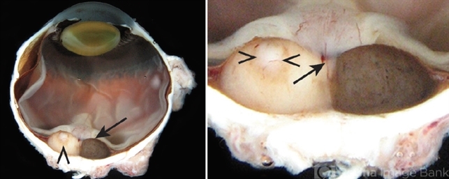
- Choroidal melanoma is often asymptomatic and diagnosis is incidental. The tumors may grow beneath the retina, or may break through the Bruch membrane and disrupt the retina. Tumors breaking through the Bruch membrane and disrupting the retina have a characteristic “mushroom” shape. The enucleation specimen in (A) shows a 2-lobed melanotic (arrow) and amelanotic (arrowhead) tumor in the posterior pole of the eye overlying the optic nerve head. In (B), higher magnification shows the optic nerve head and a feeder tumor vessel (arrow). Necrosis is present in the amelanotic tumor (arrowheads).


---thumb.jpg/image-square;max$79,0.ImageHandler "Vitrectomy Choroidal Mass")


---thumb.jpg/image-square;max$79,0.ImageHandler "unknown")

