 Initializing download.
-
 By AVIK DEY SARKAR, MS, FVRS, FAICO(VR)
Kamala Sundaram Eye Center, Chembur, Mumbai, India
Co-author(s): Dr. Naresh Babu Kannan, MS, FVRS, MBA, FASRS, Chief, Dept. of Vitreoretinal Services, Aravind Eye Hospital, Madura, India - Uploaded on Oct 31, 2024.
- Last modified by Joshua Friedman on Oct 31, 2024.
- Rating
- Appears in
- Miscellaneous
- Condition/keywords
- torpedo maculopathy, torpedo Retinopathy
- Photographer
- Dr. Avik Dey Sarkar, MBBS, MS, FVRS, FAICO, Consultant, Department of Vitreoretinal Services, Aravind Eye Hospital, Madurai, India
- Imaging device
-
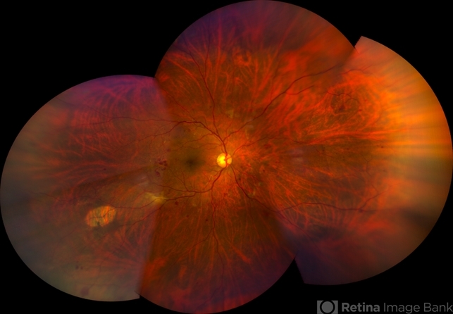
Fundus camera
Wide angled Fundus imaging with Clarus 300 - Description
- This is a 42 year old male with known history of diabetes mellitus for past 10 years. Patient presented with complains regarding presbyopia. On dilated fundoscopy, along with dot and blot hemorrhages, in the infero-temporal near-periphery outside the vascular arcade a hypopigmented torpedo-shaped lesion was noted. On OCT, outer retinal attenuation with sublesional choriocappilaris layer thinning was noted. The lesion is diagnosed as torpedo retinopathy. Torpedo maculopathy is rare in clinical practice and usually is found at the margin of temporal arcade over "Temporal Bulge". But this lesion is seen well away from the posterior pole. This case indicates the necessity of substituting the terminology "Torpedo Maculopathy" with "Torpedo Retinopathy" as mentioned earlier in ophthalmic literature.


9(Z001---thumb.BMP/image-square;max$79,0.ImageHandler "Torpedo Maculopathy")
9(Z004---thumb.BMP/image-square;max$79,0.ImageHandler "Torpedo Maculopathy - Red Free")


