Search results (64 results)
-
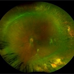
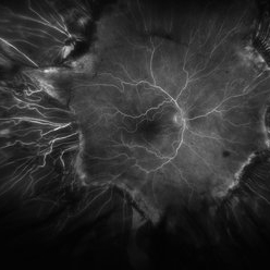 Fluorescein Angiogram of ROP With Cryo Scarring
Jul 7 2025 by Jenn Geelan
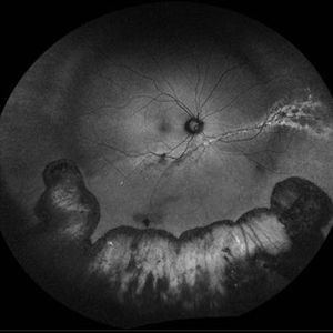
FA photo of a 34 year old male with prior stage 3 ROP with history of 360 degree cryotherapy.
Photographer: Jenn Geelan, Retina-Vitreous Surgeons of CNY
Imaging device: Optos California
Condition/keywords: cryotheraphy scar, fluorescein angiogram (FA), fundus photograph, retinopathy of prematurity (ROP), ROP, tilted disc
-
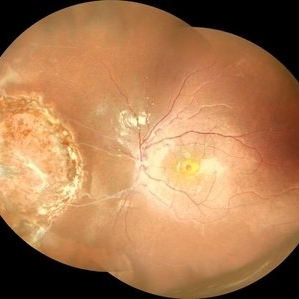
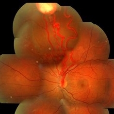
 Retinal Vasoproliferative Tumor
Jun 24 2025 by Marcelo Zas, MD PhD
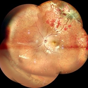
We present a case of a 33-year-old male patient, who presented with decreased visual acuity in his right eye with 20/80, presenting a primary retinal vasoproliferative tumor in the lower temporal quadrant. The tumor is associated with serous retinal detachment, hard exudation, neovascularization and telangiectasias. Lipid exudates extend toward the macula, indicating macular involvement, which may contribute to decreased visual acuity. Oi was normal with 20/20 of BCVA. The patient was treated initially with IV anti-VEGF therapy and cryotherapy.
Photographer: Marcelo Zas MD PhD
Condition/keywords: RETINAL VASOPROLIFERATIVE TUMOR
-
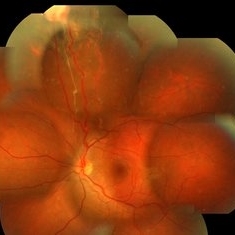
 Repaired Retinal Detachment
May 7 2025 by Kimberly Wakester
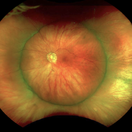
Optomap RGB montage of an 56-year-old woman with a repaired retinal detachment with scleral buckle and cryotherapy in the left eye. Patient remains stable s/p Vitreo-retinal surgery in 2007. Patient is to return in 1 year for follow up exam with repeat imaging.
Photographer: Kimberly Wakester, COA, OCT-C
Imaging device: Optos California
Condition/keywords: cryotherapy, repaired RD, scleral buckle
-
 Proliferative Sickle Cell Retinopathy
Jan 27 2025 by Virginia Gebhart
61 year-old with proliferative sickle cell retinopathy s/p cryotherapy to peripheral fibrotic NV. Eye is stable with resolving exudates and maturing cryo scar. BCVA 20/40
Photographer: Virginia Gebhart, Retina Consultants of Carolina
Imaging device: Optos California
Condition/keywords: cryotherapy, fibrotic neovascularization, sickle cell retinopathy
-
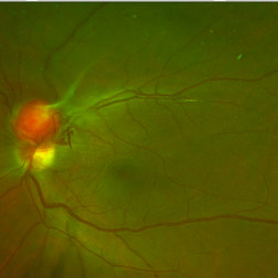
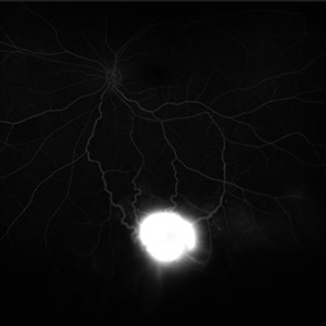 Von Hippel-Lindau Syndrome on FA
Jan 7 2025 by Jordyn Beckman
Fluorescein angiography photograph of an 37 year old female presents with reddish vascular lesion with feeder vessels for possible Von Hippel-Lindau Syndrome.
Photographer: Jordyn Beckman
Imaging device: California Optos
Condition/keywords: feeder vessel, FLUORESCEIN ANGIOGRAPHY, genetic disorder, pre-cryotherapy, Von Hippel-Lindau
-
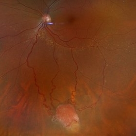 Von Hippel-Lindau Syndrome
Jan 7 2025 by Jordyn Beckman
Fundus photograph of an 37 year old female presents with reddish vascular lesion with feeder vessels for possible Von Hippel-Lindau Syndrome.
Photographer: Jordyn Beckman
Imaging device: California Optos
Condition/keywords: color fundus photograph, feeder vessel, genetic disorder, pre-cryotherapy
-
 Repaired Retinal Detachment with Multiple Breaks
Dec 9 2024 by Virginia Gebhart
FAF in 25 year old female of repaired retinal detachment 1.5 year s/p scleral buckle/cryo. Pt had been having symptoms for over a year, inferior demarcation line from retinal fluid that was present. Retina remains flat and attached under buckle. Treated lattice inferiorly, no new holes or tears. VA 20/20
Photographer: Virginia Gebhart, Retina Consultants of Carolina
Imaging device: Optos California
Condition/keywords: autofluorescence imaging, cryotherapy, demarcation line, lattice degeneration, scleral buckle
-
 Von Hippel Lindau Syndrome
Jun 9 2024 by Anjana Mirajkar, MS Ophthalmology
A widefield montage of a 23 year old female of LE case of VHL syndrome showing some hemorrhages with traction superiorly in a silicon oil filled eye with central settled retina. Cryo and laser marks are noted in periphery.
Photographer: Dr. Anjana Mirajkar -Retina Foundation, Ahmedabad
Imaging device: Mirante-Nidek
Condition/keywords: cryotherapy, exudative detachment, laser photocoagulation, vitreous hemorrhage, Von Hippel-Lindau
-
 Post-Operative Scleral Buckle
Mar 8 2024 by Ethan K Sobol, MD
The post operative week one appearance of a macula-on retinal detachment repaired with a 5mm strip encircling band, cryotherapy, and external drainage.
Photographer: Bryan Murphy, Senior Ophthalmic Photographer (Retina Group of Washington)
Imaging device: Optos California
Condition/keywords: scleral buckle
-
 Peripheral VPT Pre and Post Treatment
Aug 14 2023 by Joseph Juliano, MD
Peripheral VPT with surrounding exudation in a 35 year old woman (Top Left). Three weeks after cryotherapy there are small areas of hemorrhage along the posterior margin of the VPT (Top Right). Four months after cryotherapy, the lesion has significantly less exudation and the areas of hemorrhage have resolved (Bottom Left). Nine months after cryotherapy, there is no further exudation and the VPT is inactive with surrounding cryotherapy scarring (Bottom Right).
Condition/keywords: Vasoproliferative Tumor, VPT
-
 VPT intra-operative cryotherapy
Aug 14 2023 by Joseph Juliano, MD
Intraoperative photos of the vasoproliferative lesion before cryotherapy (left), and after triple freeze thaw cryotherapy (right). Surrounding white areas of exudation are notable around the lesion. Of note, there is tissue blue staining around the macula because this patient presented with a concurrent macular hole and an internal limiting membrane drape was performed.
Condition/keywords: Vasoproliferative Tumor, VPT
-
 Ultra-Widefield Montage of Reattached Retina and Subretinal Fluid Blebs Following Scleral Buckling Surgery
Jun 1 2021 by Kushal S Delhiwala, MBBS, MS, FMRF,FICO, FAICO
Ultra-widefield fundus photograph of left eye of a 29-year-old male who underwent scleral buckling surgery for retinal detachment.279 silicone tire and 240 band was used. Fundus shows reattached retina with adequate buckle indentation and subretinal fluid blebs along inferior arcade and nasal to disc.
Photographer: Kushal Delhiwala, Netralaya superspeciality eye hospital, Ahmedabad, Gujarat,India
Imaging device: Optos Daytona
Condition/keywords: cryotherapy, external drainage, scleral band, scleral buckle, silicone band, silicone tire
-
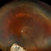
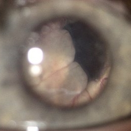
 Pupil View of Total Serous Retinal Detachment
Jan 19 2020 by Anfisa Ayalon, MD
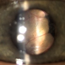
Slit-lamp photograph of a 3 -year-old male with an aggressive course of Coats' disease. Total serous retinal detachment in the right eye can be seen through the pupil. S/p laser photocoagulation, cryotherapy, retinal detachment repair with scleral buckle implantation 2 years ago. Currently, the right eye has no light perception.
Photographer: Anfisa Ayalon, MD., Meir Medical Center, Kfar Saba, Israel.
Condition/keywords: Coats' disease, pupil view, serous retinal detachment
-
 Coats' Related Total Serous Retinal Detachment
Jan 19 2020 by Anfisa Ayalon, MD
Slit-lamp photograph of a 3 -year-old male with total serous retinal detachment due to Coats' disease in the right eye. S/p laser photocoagulation, cryotherapy, retinal detachment repair with scleral buckle implantation 2 years ago. Currently, the right eye has no light perception.
Photographer: Anfisa Ayalon, MD., Meir Medical Center, Kfar Saba, Israel.
Condition/keywords: blind eye, Coats' disease, retinal detachment without retinal defect, serous retinal detachment
-
 Coats' Disease with Total Serous Retinal Detachment
Jan 19 2020 by Anfisa Ayalon, MD
Slit-lamp photograph of a 3 -year-old male with total serous retinal detachment due to Coats' disease in the right eye. S/p laser photocoagulation, cryotherapy, retinal detachment repair with scleral buckle implantation 2 years ago. Currently, the right eye has no light perception.
Photographer: Anfisa Ayalon, MD., Meir Medical Center, Kfar Saba, Israel.
Condition/keywords: chronic retinal detachment, Coats' disease, exudative retinal detachment
-
 Retinal Capillary Hemangioma
Dec 12 2019 by David L Kilpatrick, MD
OCT showing two distinct retinal capillary hemangiomas. A visually significant epiretinal membrane is also present. Work up with gene testing was negative for VHL. The plan is to proceed with PDT of the two separate lesions (half fluence for the peripapillary lesion), followed by cryotherapy / photocoagulation.
Condition/keywords: retinal capillary hemangioma
-
 Retinal Capillary Hemangioma
Dec 12 2019 by David L Kilpatrick, MD
This is a wide-field color fundus photo showing two distinct retinal capillary hemangiomas. A visually significant epiretinal membrane is also present. Work up with gene testing was negative for VHL. The plan is to proceed with PDT of the two separate lesions (half fluence for the peripapillary lesion), followed by cryotherapy / photocoagulation.
Photographer: Retina Consultants of Alabama
Imaging device: Optos
Condition/keywords: retinal capillary hemangioma
-
 Wide Field Fundus Montage of a Retinoblastoma Treated Earlier with Cryo and Laser
Aug 10 2019 by Manish Nagpal, MD, FRCS (UK), FASRS
Wide field fundus montage of a Retinoblastoma treated earlier with cryo and laser. The lesion is stable for the last five years.
Photographer: Gayathri Mohan, Retina Foundation
Imaging device: Nidek Mirante SLO
Condition/keywords: cryotherapy, laser, laser photocoagulation, retinoblastoma
-
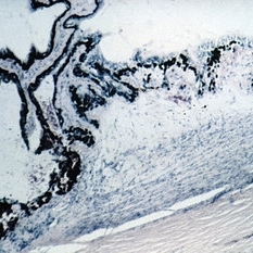 Slide 7-115
Feb 25 2019 by Lancaster Course in Ophthalmology
Cyclocryotherapy produces necrosis of the ciliary body.
Condition/keywords: ciliary, cyclocryotherapy, retinal necrosis
-
 Familial Exudative Vitreoretinopathy
Feb 2 2018 by Olivia Rainey
Ultra-wide field montage of a 37-year-old female with familial exudative vitreoretinopathy affecting her left eye. Cryotherapy, laser destruction of retinopathy, and a scleral buckle was performed to stabilize the retina in 2017.
Photographer: Olivia Rainey
Imaging device: Optos
Condition/keywords: familial exudative vitreoretinopathy (FEVR), fibrotic neovascularization, laser scarring, left eye, montage, Optos, scleral buckle, tractional retinal detachment, ultra-wide field imaging
-
 UWF of Retinal Detachment Corrected with Scleral Buckle
Aug 29 2017 by Carolyn Daley
An ultra wide field fundus photograph of a 57-year-old male who has a past history of retinal detachment corrected with scleral buckle and three treated retinal tears.
Photographer: Carolyn Daley
Imaging device: Optos Imaging
Condition/keywords: cryo-retinal tear, cryotherapy, Optos, retinal tear, scleral buckle, ultra-wide field imaging
-
 VHL With Capillary Hemangioma Post Cryo-Anti-VEGF
Dec 29 2016 by Manish Nagpal, MD, FRCS (UK), FASRS
VHL with Haemangioma status post treatment with cryo and laser.
Photographer: Rakesh Juneja
Condition/keywords: cryotherapy, hemangioma, Von Hippel-Lindau
-
 VHL With Capillary Hemangioma Pre-Rx
Dec 29 2016 by Manish Nagpal, MD, FRCS (UK), FASRS
VHL with hemangioma with feeder vessels.
Photographer: rakesh juneja
Condition/keywords: cryotherapy, hemangioma, laser, Von Hippel-Lindau
 Loading…