Search results (4 results)
-
 Von Hippel-Lindau Syndrome
Jan 7 2025 by Jordyn Beckman
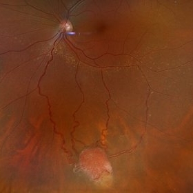
Fundus photograph of an 37 year old female presents with reddish vascular lesion with feeder vessels for possible Von Hippel-Lindau Syndrome.
Photographer: Jordyn Beckman
Imaging device: California Optos
Condition/keywords: color fundus photograph, feeder vessel, genetic disorder, pre-cryotherapy
-
 Post-Operative Scleral Buckle
Mar 8 2024 by Ethan K Sobol, MD
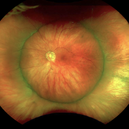
The post operative week one appearance of a macula-on retinal detachment repaired with a 5mm strip encircling band, cryotherapy, and external drainage.
Photographer: Bryan Murphy, Senior Ophthalmic Photographer (Retina Group of Washington)
Imaging device: Optos California
Condition/keywords: scleral buckle
-
 UWF of Retinal Detachment Corrected with Scleral Buckle
Aug 29 2017 by Carolyn Daley
An ultra wide field fundus photograph of a 57-year-old male who has a past history of retinal detachment corrected with scleral buckle and three treated retinal tears.
Photographer: Carolyn Daley
Imaging device: Optos Imaging
Condition/keywords: cryo-retinal tear, cryotherapy, Optos, retinal tear, scleral buckle, ultra-wide field imaging
-
 Retinal Capillary Hemangioma
Feb 10 2016 by Claudia G Hooten, MD
Fundus photo of 47-year-old male with PVR retinal detachment 2 months post cryotherapy of RCH.
Photographer: Mark D. Clark
Condition/keywords: retinal capillary hemangioma
 Loading…