Search results (64 results)
-
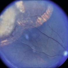
 Sudden Posterior Vitreous Detachment
Nov 9 2012 by Norman Byer
This is the appearance of the previous lesion three weeks following prophylactic cryotherapy. Continuing vitreal retinal traction has a now torn the flap completely free from the retina. The whitish cystic retinal tuft can be discerned on the upper part of the free operculum. Along the lower half of the operculum superimposed over the dark shadow of the scleral indentation one may observe numerous, delicate, vitreous fibrils actually attaching to the operculum.
Condition/keywords: cystic retinal tuft, free operculum, prophylactic cyrotherapy, retinal flap, scleral indentation, vitreoretinal traction, vitreous fibrils
-
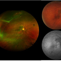
 UWF of Retinal Detachment Corrected with Scleral Buckle
Aug 29 2017 by Carolyn Daley
An ultra wide field fundus photograph of a 57-year-old male who has a past history of retinal detachment corrected with scleral buckle and three treated retinal tears.
Photographer: Carolyn Daley
Imaging device: Optos Imaging
Condition/keywords: cryo-retinal tear, cryotherapy, Optos, retinal tear, scleral buckle, ultra-wide field imaging
-
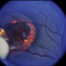
 Retinal Break at Site of Lattice Degeneration with Scleral Indentation
Nov 9 2012 by Norman Byer
This is the same case as the previous photograph. With scleral indentation slightly more posterior, the flap is seen to be associated with a large retinal tear. This is a tractional tear and it is possible that in this case the cryotherapy itself may have increased the vitreoretinal traction at this site and in this way led to this new tear. The age of the tear is unknown because it was asymptomatic, and even though the eye is aphakic the tear has not caused a clinical retinal detachment.
Condition/keywords: retinal flap, scleral indentation, tractional retinal tear, vitreoretinal traction
-
 Scleral Buckle and Cryo Color
Dec 29 2012 by Barbara Parolini, MD
Panoramic fundus photograph of a 55-year-old man after episcleral sugary for retinal detachment. An encircling scleral buckle and a superotemporal cryotherapy scar are visible.
Photographer: Barbara Parolini, MD
Imaging device: Daytona
Condition/keywords: scleral buckle
-
 Subclinical Retinal Detachment
Nov 9 2012 by Norman Byer
This 50-year-old man was treated with cryotherapy for two tiny non tractional round holes which had produced a small subclinical retinal detachment at 7:00 o’clock in this eye. Two years later he was seen with this large horseshoe tractional tear just anterior to the treated area and we must assume that it was a complication of that treatment.
Condition/keywords: cryotherapy, non-tractional holes, tractional retinal tear
-
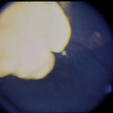
 Sudden Posterior Vitreous Detachment
Nov 9 2012 by Norman Byer
This is the same eye that was described in slide pairs 15 and 16 and shows a large tractional symptomatic retinal tear at 12 o’clock. It was caused by a posterior vitreous detachment which placed sudden traction on a cystic retinal tuft. The whitish tuft is barely visible on the flap because it is not in focus. This tear was treated successfully with cryotherapy. The next slide pair is a postoperative view of the same lesion.
Condition/keywords: cystic retinal tuft, posterior vitreous detachment, tractional retinal tear
-
 Scleral Buckle and Cryoptherapy Scar
Dec 29 2012 by Barbara Parolini, MD
Panoramic autofluorescence photograph of a 55-year-old man after episcleral sugary for retinal detachment. An encircling scleral buckle, a superotemporal cryotherapy scar and the demarcation line of hyperautofluorescence in the previous detachment area are visible.
Photographer: Barbara Parolini, MD
Imaging device: Daytona
Condition/keywords: scleral buckle
-
 Retinoblastoma Type 2 Regression After Chemo and Laser
Apr 17 2014 by Susanna S. Park, MD, PhD
Retcam fundus photograph of a 2-year-old boy with history of bilateral Group D retinoblastoma completing 6 cycles of systemic chemotherapy and retinal laser and cryotherapy with residual regressing posterior pole tumor showing predominantly type 2 regression. Pigmented rim shows scarring from prior diode and argon laser treatments.
Photographer: Ellen Redenbo, University of California Davis Eye Center
Condition/keywords: retina, retinoblastoma, type 2 regression
-
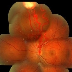
 Retinal Folds After Surgery
Jun 23 2016 by Andrea Arriola-Lopez, MD MSc
45-year-old man with history of rhegmatogenous retinal detachment and segmental scleral buckle from MIX to MXII, SF6 and cryotherapy on right eye was performed. Radial folds on indentation was seen after surgery. Three weeks later, inferior macular folds was found. The patient was asymptomatic. Observation was decided. Retina remains attach. On top, close up to macular area shows inferior folds far from fovea. Bottom, red free photograph shows no RPE changes on the same retina fold area.
Photographer: Andrea E. Arriola-López MD MSc
Imaging device: OPTOS
Condition/keywords: macular fold, retina surgery, scleral buckle
-
 Lattice Lesion
Nov 9 2012 by Norman Byer
This 55-year-old woman had had a cataract extraction five years earlier and also cryotherapy of some but not all of her lattice lesions. She was found to have this large retinal flap in the periphery near an area where cryotherapy had been applied. The next slide pair shows a different view of this lesion.
Condition/keywords: cataract extraction, cryotherapy, lattice lesion, retinal flap
-
 Retinal Capillary Hemangioma
Feb 10 2016 by Claudia G Hooten, MD
Fundus photo of 47-year-old male with PVR retinal detachment 2 months post cryotherapy of RCH.
Photographer: Mark D. Clark
Condition/keywords: retinal capillary hemangioma
-
 Retinoblastoma Recurrent Seeding After Chemotherapy
Apr 17 2014 by Susanna S. Park, MD, PhD
Retcam fundus photograph of a 2-year-old boy with history of bilateral Group D retinoblastoma completing 6 cycles of systemic chemotherapy and retinal laser and cryotherapy who was noted with recurrent peripheral seeding of tumor.
Photographer: Ellen Redenbo, University of California Davis Eye Center
Condition/keywords: retinoblastoma, tumor seeding
-
 Horseshoe Tear
Jun 24 2015 by Andree Henaine-Berra, MD
Photograph of the right eye of a 58-year-old male patient with a retinal detachment due to a peripheral horseshoe tear, showing the moment when cryotherapy is applied during the scleral bluckling procedure.
Photographer: Jorge Morales, MD. Hospital General "Dr. Manuel Gea Gonzalez". Mexico City.
Condition/keywords: acute retinal detachment, cryotherapy, scleral buckle
-
 Group D Retinoblastoma After Chemo and Laser
Apr 17 2014 by Susanna S. Park, MD, PhD
Retcam fundus photograph of a 2 year old boy with history of bilateral Group D retinoblastoma completing 6 cycles of systemic chemotherapy and retinal laser and cryotherapy with residual regressing posterior pole tumor showing type 3 (type 1 and 2) regression pattern. Some pigmented scarring and hemorrhage are also noted nasal to the disc from prior laser treatment of tumor.
Photographer: Ellen Redenbo
Condition/keywords: retina
-
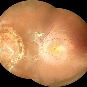
 Coat's Disease with Cryotherapy
Aug 14 2013 by Jason S. Calhoun
Young male with Coat's disease with cryotherapy in the right eye. VA was 20/20, right eye. Left eye was completely normal. Patient will be followed up in 3-months
Photographer: Jason S. Calhoun, Department of Ophthalmology, Mayo Clinic Jacksonville, Florida
Imaging device: TOPCON TRC 50-EX
-
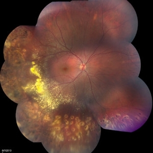
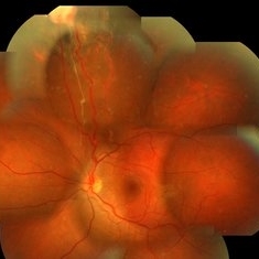
 Familial Exudative Vitreoretinopathy
Feb 2 2018 by Olivia Rainey
Ultra-wide field montage of a 37-year-old female with familial exudative vitreoretinopathy affecting her left eye. Cryotherapy, laser destruction of retinopathy, and a scleral buckle was performed to stabilize the retina in 2017.
Photographer: Olivia Rainey
Imaging device: Optos
Condition/keywords: familial exudative vitreoretinopathy (FEVR), fibrotic neovascularization, laser scarring, left eye, montage, Optos, scleral buckle, tractional retinal detachment, ultra-wide field imaging
-
 Retinal Detachment Repaired With Scleral Buckle
Oct 2 2013 by Jerald A. Bovino, MD
A retinal detachment was repaired with an encircling scleral buckle. Nasally a cryotherapy scar is visible.
Condition/keywords: cryotheraphy scar, encircling scleral buckle
-
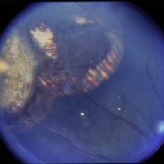
 VHL With Capillary Hemangioma Pre-Rx
Dec 29 2016 by Manish Nagpal, MD, FRCS (UK), FASRS
VHL with hemangioma with feeder vessels.
Photographer: rakesh juneja
Condition/keywords: cryotherapy, hemangioma, laser, Von Hippel-Lindau
-
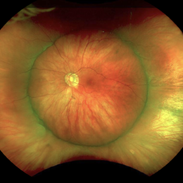
 Wide Field Fundus Montage of a Retinoblastoma Treated Earlier with Cryo and Laser
Aug 10 2019 by Manish Nagpal, MD, FRCS (UK), FASRS
Wide field fundus montage of a Retinoblastoma treated earlier with cryo and laser. The lesion is stable for the last five years.
Photographer: Gayathri Mohan, Retina Foundation
Imaging device: Nidek Mirante SLO
Condition/keywords: cryotherapy, laser, laser photocoagulation, retinoblastoma
-
 VHL With Capillary Hemangioma Post Cryo-Anti-VEGF
Dec 29 2016 by Manish Nagpal, MD, FRCS (UK), FASRS
VHL with Haemangioma status post treatment with cryo and laser.
Photographer: Rakesh Juneja
Condition/keywords: cryotherapy, hemangioma, Von Hippel-Lindau
-
 Retinoblastoma
Dec 5 2014 by David Callanan, MD
2-year-old black male, retinoblastoma, full term infant, normal delivery; S/P enucleation OS, S/P cryotherapy OD x 3 ; VA fixes & follows OD.
Condition/keywords: retinoblastoma
-
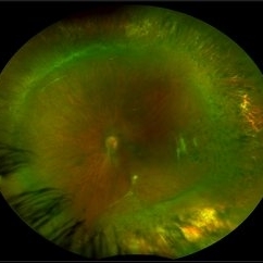
 Post-Operative Scleral Buckle
Mar 8 2024 by Ethan K Sobol, MD
The post operative week one appearance of a macula-on retinal detachment repaired with a 5mm strip encircling band, cryotherapy, and external drainage.
Photographer: Bryan Murphy, Senior Ophthalmic Photographer (Retina Group of Washington)
Imaging device: Optos California
Condition/keywords: scleral buckle
-
 Retinoblastoma
Dec 5 2014 by David Callanan, MD
2-year-old black male, retinoblastoma, full term infant, normal delivery; S/P enucleation OS, S/P cryotherapy OD x 3 ; VA fixes & follows OD.
Condition/keywords: retinoblastoma
-
 Retinoblastoma
Dec 5 2014 by David Callanan, MD
2-year-old black male, retinoblastoma, full term infant, normal delivery; S/P enucleation OS, S/P cryotherapy OD x 3 ; VA fixes & follows OD.
Condition/keywords: retinoblastoma
-
 Retinoblastoma
Dec 5 2014 by David Callanan, MD
2-year-old black male, retinoblastoma, full term infant, normal delivery; S/P enucleation OS, S/P cryotherapy OD x 3 ; VA fixes & follows OD.
Condition/keywords: retinoblastoma
 Loading…