Search results (11 results)
-
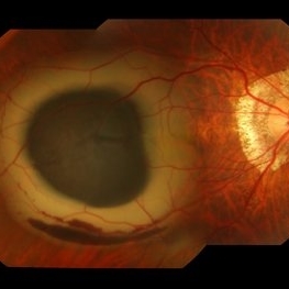
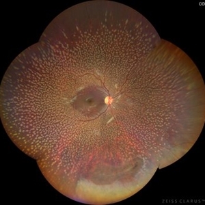 Benign Familial Fleck Retina
Dec 21 2023 by Vishal Agrawal, MD, FRCS,FACS,FASRS
10-year male with high myopia on examination revealed diffuse flecks distributed all over fundus in both eyes sparing macula. Inferior lattice with WWOP areas were also noted in right eye.
Photographer: Dr Ayushi
Imaging device: Clarus 700
Condition/keywords: fleck retinopathy, myopia
-
 Myelinated Nerve Fibre (MNF)
Jun 17 2023 by Harsh Vardhan Singh, MS
Fundus photograph of 32-year-old male having good best corrected visual acuity in both eyes with right eye having high myopia & MNF as incidental finding
Photographer: Dr Harsh Vardhan Singh, Assistant Professor, AIIMS, Guwahati
Condition/keywords: medullated nerve fibers, MNF, myelinated nerve fiber layer, myelinated nerve fibers, Nerve fiber layer arrangements, NFL
-
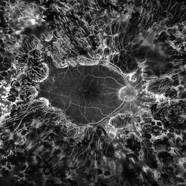
 Multifocal Choroiditis and Panuveitis- Schlaegel lines
Nov 16 2021 by Manuel Ángel Alcántara Delgado, MD
Optomap ultra-widefield retinal imaging of an 52-year-old woman showed multiple punched-out chorioretinal lesions and 2 rows of peripheral curvilinear pigmented chorioretinal streaks (Schlaegel lines).
Photographer: Manuel Ángel Alcántara Delgado. Conde de Valenciana.
Condition/keywords: multifocal choroiditis, myopia, retina, uveitis
-
 PDR; High Myopia; PRP
May 2 2019 by Carissa Hurdstrom
PDR; high myopia; PRP
Imaging device: Optos
Condition/keywords: fluorescein angiogram (FA), high myopia, pan-retinal photocoagulation (PRP), proliferative diabetic retinopathy (PDR)
-
 Optos Picture With Speculum: Dislocated Natural Lens
Oct 9 2018 by John S. King, MD
55-year-old white female with history of pathologic myopia+, lattice (laser), SB OU (1990s), and dislocated natural lenses OU that had been watched for years. In the fellow eye she developed phacolytic glaucoma and a PPV, PPL was performed. Plan for both eyes are monitoring. I wanted to get a good picture of her lens today with the optos machine, as the other pics had artifact from the lower lid. It worked out well to use a speculum in the left eye. Vision cc is 20/400 J1+ OD and 20/40 J2 OS; aphakic OU; vitreous clear OD; dislocated lens OS (see pic); retinas attached.
Photographer: Maisee Yang
Imaging device: Optos California
Condition/keywords: dislocated crystalline lens, pathologic myopia, scleral buckle, staphyloma
-
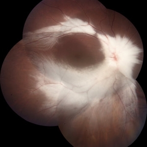
 Giant Retinal Tear-Retinal Detachment
May 25 2017 by Somnath Chakraborty, MD
Fundus photograph of a 45-year-old female with pathological myopia and retinal detachment secondary to giant retinal tear.
Photographer: Saptarshi Mehta, Retina Institute of Bengal
Condition/keywords: giant retinal tear, retinal tear
-
 IOL With BAG in Vitreous of Myopic Eye
Apr 14 2017 by Manish Nagpal, MD, FRCS (UK), FASRS
50-year-old male having myopia presented with a IOL in vitreous within its bag.
Photographer: Pooja Barot
Condition/keywords: intraocular lens (IOL), intraocular lense in vitreous, intraocular lense with bag, myopia
-
 Asymptomatic Superior Retinal Detachment
May 5 2016 by Steven J Ryder, MD
38-year-old African American female with moderate myopia (-4.50 Sph OU) and asymptomatic superior retinal detachment in the right eye. Montage fundus photography showing localized retinal detachment superiorly with single full-thickness retinal break at 12:00.
Photographer: Luis Bernhard, Miami VA Healthcare System
Imaging device: Topcon
Condition/keywords: asymptomatic, full thickness retinal hole, myopia, retinal break, retinal detachment with retinal defect
-
 Submacular Hemorrhage
Mar 12 2016 by Sjakon G Tahija, MD
This a fundus photograph of a high myope who presented with a submacular hemorrhage.
Photographer: Avris Siahaan, Klinik Mata Nusantara, Jakarta, Indonesia
Condition/keywords: high myopia, spontaneous submacular hemorrhage
-
 Foveoschisis secondary to high myopia
Mar 13 2015 by Niloofar Piri, MD
Infrared and HD-OCT of the right eye in a 55-year-old African American female with high myopia (more than -6.00 D), BCVA: 20/25 OU Cartwheel appearance of the fovea in the infrared imaging is visible. HD- OCT demonstartes schisis in different layers of the retina (both NFL and OPL; notice stretching of the Muller cells); VMT is also present . Outer retinal layers are preserved which explains the good vision . She had the same findings in OS.
Photographer: Niloofar Piri, MD
Imaging device: Heidelberg Spectralis
Condition/keywords: high myopia, retinoschisis
-
 OCT Myopic Staphyloma With Schisis and ERM
Apr 24 2014 by Scott E. Pautler, MD
OCT of high myope with asymptomatic macular schisis.
Imaging device: Heidelberg Spectralis
Condition/keywords: foveal schisis, maculopathy, maculoschisis, optical coherence tomography (OCT), pathologic myopia, staphyloma
 Loading…