Search results (317 results)
-
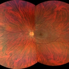
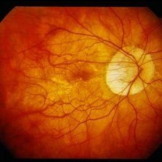
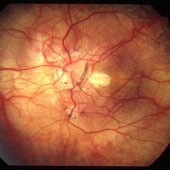
 Lacquer Cracks in Pathologic Myopia
Apr 19 2013 by Theodore Leng, MD, MS, FASRS
Color fundus photograph of a 48-year-old woman with lacquer cracks in the setting of pathologic myopia.
Condition/keywords: lacquer cracks, pathologic myopia
-
 Tigroid Fundus
Aug 31 2021 by Ricardo Leitão Guerra
True color (RGB) confocal scanning laser ophthalmoscopy of a 37-year-old male with myopia highlighting choroidal vessels and vortex veins.
Photographer: Juliana Rio, MD. Leitão Guerra - Oftlamologia, Salvador - Brazil
Imaging device: Zeiss Clarus 7000
Condition/keywords: myopia, retina, tigroid fundus, vortex vein
-
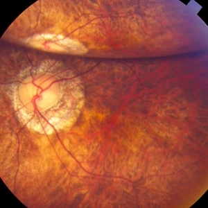
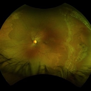
 Inferior Rhegmatogenous Retinal Detachment with Subretinal Fibrosis
Aug 23 2012 by Gabriela Lopezcarasa Hernandez, MD
Asymptomatic 25-year-old woman with high myopia.
Photographer: Gabriela Lopezcarasa Hernandez, Hospital Angeles Lomas
Imaging device: FF4
Condition/keywords: high myopia, subretinal fibrosis
-
 "Internal Mirroring" Effect by Intraocular Gas
Mar 25 2014 by Homayoun Tabandeh, MD, FASRS
"Internal mirroring" by residual intraocular gas in a highly myopic patient 3 weeks post repair of retinal detachment with pars plana vitrectomy and C3F8 gas.
Photographer: Danny Rivas
Condition/keywords: high myopia, intraocular gas
-
 Myopic CNV
Jan 11 2013 by Alex P. Hunyor, MD
Myopic macular degeneration complicated by subretinal neovascularisation, left eye.
Condition/keywords: high myopia, myopia, myopic choroidal neovascularization (CNV)
-
 Myopic Choroidal Neovascular Membrane
Mar 25 2013 by Ratimir Lazic, MD, PhD
Color fundus photography of a 33-year-old female. In macular area subretinal hemorrhage can be seen. Area of atrophy temporal from PNO. Myopic changes of posterior pole and mid periphery can be noticed. The patient has been treated with 2 consecutive ranibizumab intravitreal injections. BCVA at baseline was 0,05 (Snellen lines) and 0,3 (Snellen lines) 2 months after.
Photographer: Marko Lukic, MD
Imaging device: Zeis Visucam Lite 2
Condition/keywords: high myopia, myopic choroidal neovascularization (CNV), ranibizumab
-
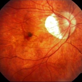
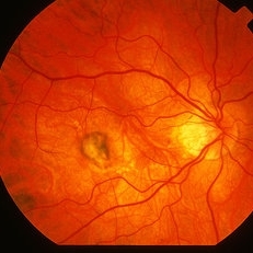
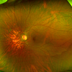
 Peripapillary Atrophy With High Myopia
Feb 4 2015 by H. Michael Lambert, MD
Peripapillary atrophy and central macular degeneration seen in high myopia.
Condition/keywords: high myopia, peripapillary atrophy
-
 Fuch's Spot
Apr 2 2019 by Gary R. Cook, MD, FACS
20-year-old patient with high myopia and a Fuch's spot OD.
Condition/keywords: Fuchs, high myopia, pathologic myopia
-
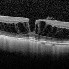
 OCT Myopic Staphyloma With Schisis and ERM
Apr 24 2014 by Scott E. Pautler, MD
OCT of high myope with asymptomatic macular schisis.
Imaging device: Heidelberg Spectralis
Condition/keywords: foveal schisis, maculopathy, maculoschisis, optical coherence tomography (OCT), pathologic myopia, staphyloma
-
 Giant Retinal Tear
Oct 9 2012 by Audina M. Berrocal, MD FASRS
Teenager with high myopia and a GRT
Photographer: Ditte Hess CRA, BPEI
Imaging device: Fundus Camera
Condition/keywords: high myopia, retinal degeneration, retinal tear
-
 Peripapillary Atrophy With High Myopia
Feb 4 2015 by H. Michael Lambert, MD
Peripapillary atrophy and central macular degeneration seen in high myopia.
Condition/keywords: high myopia, peripapillary atrophy
-
 Myopic Macular Schisis with Lamellar Macular Hole
May 26 2014 by John T. Thompson, MD
Spectral domain OCT of patient with high myopia and myopic macular schisis resulting in lamellar macular hole.
Condition/keywords: lamellar macular hole, myopic macular schisis
-
 Lacquer Cracks in Pathologic Myopia
Apr 19 2013 by Theodore Leng, MD, MS, FASRS
Red free fundus photograph of a 48-year-old woman with lacquer cracks in the setting of pathologic myopia.
Condition/keywords: lacquer cracks, pathologic myopia
-
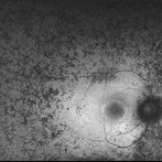 Retinitis Pigmentosa - Autofluorescence OD
Jun 18 2018 by Hosam Attia, MD
Ultra-wide fundus auto-fluorescence photograph of a 38-year-old African, American female with degenerative myopia, unilateral RP variant, depicting extensive mid-peripheral bone spicules hypo-autofluorescence, extending further into the periphery w/ relative sparing of the macula OD VF 30-V showed severe peripheral constriction OD, enlarged BS OS and OCT showed severe ellipsoid zone degeneration with saucerization and cystoid macular degeneration with no obvious late macular leakage on FA (Both, not shown)
Imaging device: Optos California
Condition/keywords: bone spicule, peripheral bone spicules, retinitis pigmentosa
-
 Myopic Choroidal Neovascularization
Aug 23 2012 by Gabriela Lopezcarasa Hernandez, MD
19-year-old male who complains of scotoma and metamorphopsias.
Photographer: Gabriela Lopezcarasa Hernandez, Macular Retina Consultores
Imaging device: Heidelberg Spectralis
Condition/keywords: choroidal neovascularization (CNV), myopia
-
 Optos Picture With Speculum: Dislocated Natural Lens
Oct 9 2018 by John S. King, MD
55-year-old white female with history of pathologic myopia+, lattice (laser), SB OU (1990s), and dislocated natural lenses OU that had been watched for years. In the fellow eye she developed phacolytic glaucoma and a PPV, PPL was performed. Plan for both eyes are monitoring. I wanted to get a good picture of her lens today with the optos machine, as the other pics had artifact from the lower lid. It worked out well to use a speculum in the left eye. Vision cc is 20/400 J1+ OD and 20/40 J2 OS; aphakic OU; vitreous clear OD; dislocated lens OS (see pic); retinas attached.
Photographer: Maisee Yang
Imaging device: Optos California
Condition/keywords: dislocated crystalline lens, pathologic myopia, scleral buckle, staphyloma
-
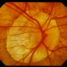
 Myopic Shift With Tilted Optic Disc.
Jul 11 2013 by Jason S. Calhoun
Black female who has a myopic shift shows tilted optic disc in the right eye.
Photographer: Jason S. Calhoun, Department of Ophthalmology, Mayo Clinic Jacksonville, Florida
Condition/keywords: high myopia
-
---thumb.jpg/image-square;max$300,300.ImageHandler) Birdshot Choroidopathy
Birdshot Choroidopathy
Oct 9 2013 by Maurice F. Rabb
Forty two year old white female first noted flashing lights in her left eye at the age of 30. Although she had many previous eye examinations for low grade myopia, she had never had a dilated fundus examination. The evaluation twelve years ago disclosed 20/20 acuity in each eye with a myopic correction, an afferent pupillary defect on the left, no evidence of anterior segment inflammation in either eye, a full field on the right and markedly constricted field on the left, fundus pigmentary abnormalities extending beyond the equator in each eye, and narrow vessels with pigment migration into the retina in the left eye only.
Condition/keywords: birdshot choroidopathy
-
 Myopic CNV
May 2 2013 by Henry J. Kaplan, MD
Subretinal membrane in high myopia.
Condition/keywords: myopic choroidal neovascularization (CNV)
-
 Progressive Bifocal Chorioretinal Atrophy
Feb 1 2015 by Andree Henaine-Berra, MD
Fundus photograph of the left eye of an 13-year-old female patient with poor vision, high myopia and nystagmus. The image shows macular dragging, a limited area of chorioretinal atrophy temporal to the optic disc and an extense area of chorioretinal atrophy temporal to the macula that extended to the extreme periphery.
Photographer: Andree Henaine-Berra, MD
Condition/keywords: chorioretinal atrophy
-
 Dome-Shaped Macula With Subretinal Fluid
Jun 14 2018 by Gerardo Garcia-Aguirre, MD
EDI OCT of the right eye of a 17-year-old highly myopic girl. Subfoveal fluid is present. There is choroidal thinning, and scleral thickening in the foveal area.
Photographer: Gerardo Garcia-Aguirre, MD
Imaging device: Heidelberg Spectralis
Condition/keywords: dome shaped macula, high myopia
-
 High Myopia
May 2 2013 by Henry J. Kaplan, MD
Chorioretinal atrophy in high myopia and tilted disc.
Condition/keywords: high myopia, tilted disc
-
---thumb.jpg/image-square;max$300,300.ImageHandler) Retinectomy With Diathermy in a Giant Tear
Retinectomy With Diathermy in a Giant Tear
Mar 13 2014 by Marcelo Zas, MD PhD
The image show a giant tear in a myopic patient. We use diathermy to avoid intraop bleeding.
Photographer: Marcelo Zas MD PhD
Condition/keywords: giant retinal tear, myopia
-
 Myopia with Lattice Degeneration and White Without Pressure in the Setting of Marfan's Syndrome
Aug 31 2020 by Sophia El Hamichi, MD
A 1-year-old female with Marfan's syndrome, myopia OU, congenital nystagmus and exotopia OD. Ultra-wide field imaging of her left eye showed lattice degeneration with atrophic retinal holes temporally, in addition to multiple sections of white without pressure.
Imaging device: Optos
Condition/keywords: atrophic retinal hole, lattice degeneration, Marfan's syndrome, myopia, Optos, ultra-wide field imaging
-
 White Without Pressure
Mar 13 2020 by Anfisa Ayalon, MD
Fundus photograph of a 26-year-old woman with high myopia. Note inferotemporally margins of sharply demarcated WWP area.
Photographer: Anfisa Ayalon, MD., Meir Medical Center, Kfar Saba, Israel.
Imaging device: California, Optos 200 DTX
Condition/keywords: myopia, white without pressure
 Loading…