Search results (317 results)
-
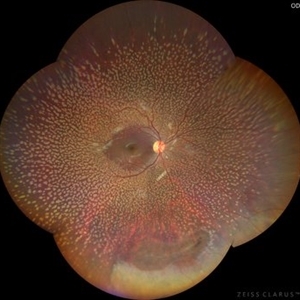
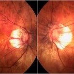
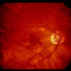
 Asymptomatic Superior Retinal Detachment
May 5 2016 by Steven J Ryder, MD
38-year-old African American female with moderate myopia (-4.50 Sph OU) and asymptomatic superior retinal detachment in the right eye. Montage fundus photography showing localized retinal detachment superiorly with single full-thickness retinal break at 12:00.
Photographer: Luis Bernhard, Miami VA Healthcare System
Imaging device: Topcon
Condition/keywords: asymptomatic, full thickness retinal hole, myopia, retinal break, retinal detachment with retinal defect
-
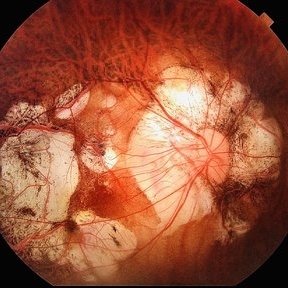
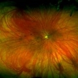
 Benign Familial Fleck Retina
Dec 21 2023 by Vishal Agrawal, MD, FRCS,FACS,FASRS
10-year male with high myopia on examination revealed diffuse flecks distributed all over fundus in both eyes sparing macula. Inferior lattice with WWOP areas were also noted in right eye.
Photographer: Dr Ayushi
Imaging device: Clarus 700
Condition/keywords: fleck retinopathy, myopia
-
 Bilateral Extreme Myopia
May 2 2024 by Matias Iglicki, MD
Extreme myopic stage
Photographer: Matias Iglicki
Imaging device: DAYTONA
Condition/keywords: myopia
-
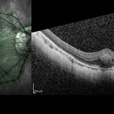 Central Bouquet Hemorrhage
May 31 2025 by Moazzam Parvez
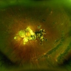
OCT image of a 26 year old gentleman of right eye macula with a central foveolar cotton ball like lesion . Inward traction by Müller cells over CB causes upward displacement of foveal cones without major disturbance of ellipsoid zone (EZ) and ELM . Cotton ball sign is characterized by small, fuzzy subfoveal hyperreflective area between the inner segment ellipsoid zone (EZ) and the interdigitation zone (IZ) .
Photographer: Dr Moazzam Parvez , Netralayam , Kolkata
Imaging device: Heidelberg Spectralis
Condition/keywords: Central bouquet haemorrhage, macula, myopia
-
 Degenerative Myopia
Apr 21 2024 by César Adrián Gómez Valdivia, MD
Degenerative Myopia
Photographer: Erika Paulina Ornelas Cazares
Imaging device: Topcon TRC-50 DX
Condition/keywords: degenerative myopia, myopia
-
 EDI OCT - Dome Shaped Macula in a Myopic Patient
Jan 24 2015 by Roy Schwartz, MD
Dome shaped macula in an 80-year-old man with myopic staphyloma. The EDI showed thickened choroid and sclera.
Photographer: Galit Yair Pur
Condition/keywords: enhanced depth imaging, myopia, optical coherence tomography (OCT)
-
 High Myopia
Dec 7 2019 by Anfisa Ayalon, MD
Fundus photograph of a 55-year-old woman with high myopia.
Photographer: Anfisa Ayalon,MD., Meir Medical Center, Kfar Saba, Israel.
Condition/keywords: high myopia, myopia, peripapillary atrophy
-
 High Myopia with Cobblestone Degeneration
Nov 5 2019 by Nichole Lewis
50-year-old female with high myopia, diffuse myopic thinning and cobblestone degeneration.
Photographer: Nichole Lewis
Imaging device: Optos
Condition/keywords: high myopia, myopia, paving stone degeneration
-
 High Myopia with Lacquer Cracks
Apr 2 2019 by Gary R. Cook, MD, FACS
High myopia with lacquer cracks; -12.25D
Condition/keywords: high myopia, lacquer cracks, myopia
-
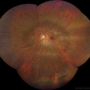 High Myopia with Posterior staphyloma
Nov 7 2023 by Harsh Vardhan Singh, MS
27-year old with both eyes high myopia & posterior staphyloma with left eye peripheral lattice degeneration & white without pressure
Photographer: Harsh Vardhan Singh
Imaging device: Clarus 700
Condition/keywords: lattice degeneration, myopia, peripheral lattice degeneration, posterior staphylomaloma, white without pressure
-
 Horseshoe Tear
Apr 9 2017 by Aliya Sultana
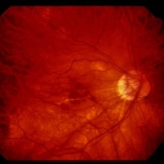
Fundus photograph of an 41-year-old man with multiple horseshoe tears in the retina, patient is myopic with -3.00 Dsph in both eyes. Suddenly patient noticed loss of vision in left eye , presented to our department next day. Other eye showed lattice degeneration . Patient underwent pars plana vitrectomy with silicone oil tamponade.
Photographer: Dr Aliya Sultana , Assistant Professor,Sarojini Devi Eye Hospital, Hyderabad, Telangana. India.
Condition/keywords: myopia
-
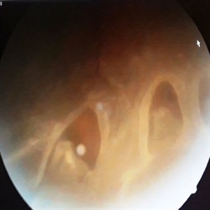
 IOL With BAG in Vitreous of Myopic Eye
Apr 14 2017 by Manish Nagpal, MD, FRCS (UK), FASRS
50-year-old male having myopia presented with a IOL in vitreous within its bag.
Photographer: Pooja Barot
Condition/keywords: intraocular lens (IOL), intraocular lense in vitreous, intraocular lense with bag, myopia
-
 LARGE OPERCULATED HOLE
Oct 16 2023 by ANKIT JAIN
Left Eye Montage of 50 years old male with high myopia with large operculated hole
Photographer: DR ANKIT JAIN
Imaging device: MIRANTE
Condition/keywords: High Myopia, myopia, operculated retinal hole
-
Macular hole in myopic patient
Nov 4 2022 by Manish Nagpal, MD, FRCS (UK), FASRS
This is a case earlier operated three times for myopic retinal detachment. Buckling was done followed by vilion oil injection and then silicon oil removal, now the patient developed a macular hole. Staining with brilliant blue followed by ILM peeling with a longer length forceps was carried out. Air fluid exchange with drainage over the hole and gas injection was done
Condition/keywords: brilliant blue staining, ILM peeling, myopia, myopic macular hole, video, vitrectomy
-
 Multifocal Choroiditis and Panuveitis- Schlaegel lines
Nov 16 2021 by Manuel Ángel Alcántara Delgado, MD
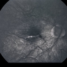
Optomap ultra-widefield retinal imaging of an 52-year-old woman showed multiple punched-out chorioretinal lesions and 2 rows of peripheral curvilinear pigmented chorioretinal streaks (Schlaegel lines).
Photographer: Manuel Ángel Alcántara Delgado. Conde de Valenciana.
Condition/keywords: multifocal choroiditis, myopia, retina, uveitis
-
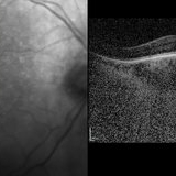
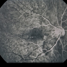
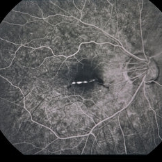
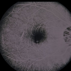
 Myope With Staphyloma and Vitreous Detachment
Jun 11 2016 by Philip J. Polkinghorne, MD
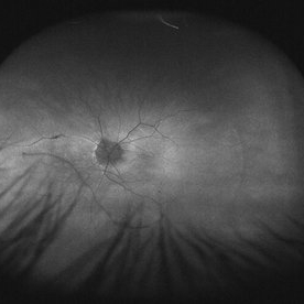
Fundus autofluorescence of a myope with PVD and staphyloma.
Imaging device: Optos FAF
Condition/keywords: degenerative myopia, myopia, staphyloma
-
 Myopia
-
 Myopia
-
 Myopia
-
 Myopia
-
 Myopia
-
 Myopia
-
 Myopia
-
 Myopia
-
 Myopia
 Loading…