File number: 27206
Comments
-
 Manish Nagpal, MD, FRCS (UK), FASRS (October 5 2017)
Fantastic image somnath :)
-
 Suber S. Huang, MD, MBA, FASRS (July 28 2017)
Difficult shot and difficult case. Thanks! Best imaged if the retina is in focus and choroid in the background.
-
 Deepak Bhojwani, MS (July 1 2017)
CURTAIN FALLS!!!!
Sign in to comment.
 Initializing download.
-
 By Somnath Chakraborty, MD
Retina Institute of Bengal, Siliguri
Co-author(s): Pulok Roy, Retina Institute of Bengal - Uploaded on May 25, 2017.
- Last modified by Caroline Bozell on Sep 28, 2017.
- Image of the week
-
Oct 1, 2017
View all images of the week - Rating
- Appears in
- Miscellaneous
- Condition/keywords
- retinal tear, giant retinal tear
- Photographer
- Saptarshi Mehta, Retina Institute of Bengal
- Imaging device
- Fundus camera
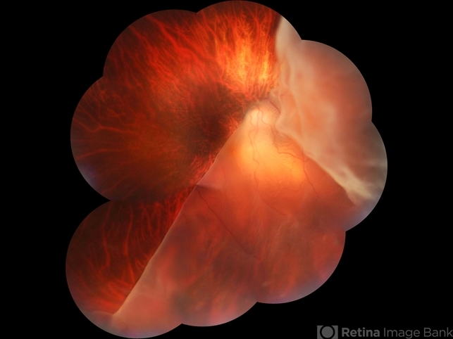
- Description
- Fundus photograph of a 45-year-old female with pathological myopia and retinal detachment secondary to giant retinal tear.


---thumb.jpg/image-square;max$79,0.ImageHandler "Cryo-Retinal Tear")
---thumb.JPG/image-square;max$79,0.ImageHandler "Giant retinal tear")


