Search results (13 results)
-
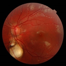
 Presumed Congenital Toxoplasmosis
Aug 16 2025 by Vishal Agrawal, MD, FRCS,FACS,FASRS
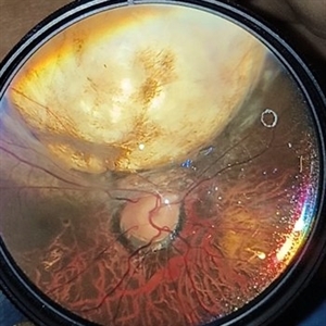
Fundus picture of 7 a year-old boy with esotropia. OCT showed complete atrophy & disorganization of the overlying RPE and neurosensory retina.
Photographer: Dr Ayushi Gupta
Imaging device: Clarus 700
Condition/keywords: coloboma of macula, toxoplasmosis
-
 Fundal Coloboma
Mar 6 2023 by Kalyan Singh
34 year old male with fundal Coloboma presented for refractive correction.
Photographer: Kalyan Singh, GSVM medical college, Kanpur
Imaging device: Smartphone (1 plus 10R)
Condition/keywords: coloboma
-
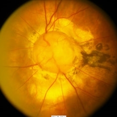
 Chorioretinal coloboma involving disc and macula
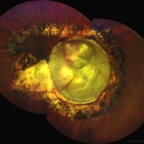
Mar 21 2022 by T. P . VIGNESH, MBBS,MS
Fundus photo of Right eye of a 55 year male patient revealing a fovea sparing well barraged chorioretinal coloboma involving the disc and the macula .
Photographer: Bharathi Singaravel
Imaging device: Zeiss Clarus
Condition/keywords: chorioretinal coloboma, coloboma of optic disc
-
 Coloboma involving the Optic nerve, Retina, and Choroid
Dec 6 2021 by Jesus Lozano, MD
78-year-old woman after prophylactic laser photocoagulation (PLP) for her RE Coloboma involving the optic nerve, retina, and choroid. At 6 month follow up, patient preserved her FC vision as it was before the procedure. Retina attached.
Photographer: Yair Bet Yosef, Hadassah Medical Center. Israel
Imaging device: Optos Silverstone fundus image
Condition/keywords: coloboma, coloboma of choroid, coloboma of macula, coloboma of optic disc, PLP, prophylactic photocoagulation
-
 Morning Glory Disc Anomaly
Nov 11 2020 by Yoshihiro Yonekawa, MD, FASRS
Color fundus photograph of a young boy with morning glory disc anomaly. Notice the concavity surrounding the enlarged disc, radial vasculature, and nasally dragged macula. MRI was negative for moyamoya disease, a known association.
Photographer: Alicia Thresher, Mid Atlantic Retina
Imaging device: Topcon
Condition/keywords: disc coloboma, Morning Glory Syndrome, pediatric retina
-
 Retinal Detachment Associated with Coloboma
Aug 23 2020 by Noy Ashkenazy, MD, MS
Fundus photograph of a 2-year-old boy with a history of CHARGE syndrome. The image nicely illustrates a retinal detachment associated with a congenital coloboma.
Photographer: Giselle DeOliveira
Imaging device: Retcam III
Condition/keywords: CHARGE syndrome, chronic retinal detachment, coloboma, pediatric retina
-
 Coloboma
Oct 2 2019 by John S. King, MD
27-year-old white female with bilateral, isolated, inferior, chorioretinal colobomas; she has a history of retinal laser anterior to the edge of the coloboma OD secondary to a limited RD. This is the right eye.
Photographer: Shelly Blair
Imaging device: Optos CA
Condition/keywords: coloboma of choroid
-
 Retinochoroidal Coloboma With Aberrant Vasculature
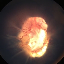
Nov 10 2018 by Chintan D Desai, MBBS, DO, DNB, FICO
Fundus photo montage of a 32-year-old female with a retinochoroidal coloboma Ida Mann classification type 3 with a spring coil shaped aberrant vessel.
Photographer: Kankan Talukdar
Imaging device: Zeiss FF4
Condition/keywords: chorioretinal coloboma
-
 Optic Disc Coloboma`
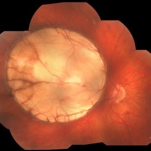
Mar 26 2018 by Purva Patwari
16-year-old female patient with vision of 6/60 presented with diminished vison. Other eye was normal.She had a normal birth history and developmental milestone. Look at the optic disc coloboma extending upto the macula. Intercalary membrane looks normal.
Photographer: Dr Purva Patwari, Patwari Retina Center, Ahmedabad, Gujarat , India
Imaging device: ZEISS VISU 500
Condition/keywords: coloboma, coloboma of optic disc, optic disc
-
 Coloboma
Jan 23 2018 by JEFFERSON R SOUSA, Tecg.º (Biomedical Systems Technology)
Male patient, 22 years old, with low vision since infancy. In retinal and retinal mapping examinations, important alterations were observed in the formation of retinochoroidal structures suggestive of coloboma.
Photographer: JEFFERSON R SOUSA - Study Center and Ophthalmological Research Dr. Andre M V Gomes, Dr. Suel Abujamra Institute São Paulo-Brazil
Imaging device: Acquisition of the image in the Camera background Topcon TRC-50 Dx - IA, Keystone field photo of 50 Degrees. Composition automatic of Imaginet with manual adjustment
Condition/keywords: coloboma, coloboma of choroid
-
 Chorio-Retinal Coloboma
Aug 4 2017 by Marco D'Angelo
Left eye, 7-year-old female patient, normal visual acuity,
Photographer: Dr. Marco D'Angelo, S.Chiara Hospital, Trento, Italy
Imaging device: Topcon TRC-NW 6S
Condition/keywords: coloboma
-
 Optic Disc Coloboma
Sep 18 2016 by John T. Thompson, MD
Optic disc coloboma
Imaging device: Zeiss FF4
Condition/keywords: coloboma, optic disc
-
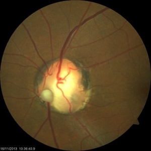 Optic Nerve Coloboma With 2 Pits, Nasal and Temporal Color
Nov 21 2013 by Alexandre Durao Alves Pereira, MD
Fundus photograph, color, red free, blue lite and FAF of a optic nerve coloboma with 2 pits, one nasal and other temporal.
Photographer: Alexandre Pereira
Imaging device: Visucam 300
Condition/keywords: color photo, optic nerve coloboma
 Loading…