Search results (277 results)
-
 Chorioretinal Coloboma
Oct 6 2025 by Seif Allah Anwar
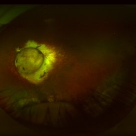
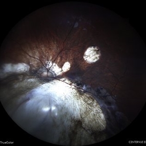
Fundus photograph of the patient left eye showing large, well-demarcated, excavated chorioretinal coloboma involving the inferior fundus, extending from the optic disc to the periphery. The lesion appears white due to bare sclera visibility, with absence of overlying choroid and retina. Retinal vessels course over the colobomatous area inferiorly.
Photographer: Dr. Seif Anwar, FRCSEd
Imaging device: Centervue Eidon
Condition/keywords: chorioretinal coloboma
-
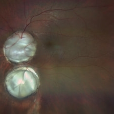
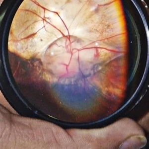
 Optic Nerve Coloboma and Rhegmatogenous Retinal Detachment
Sep 30 2025 by Píndaro Alonso Cruz-Benitez
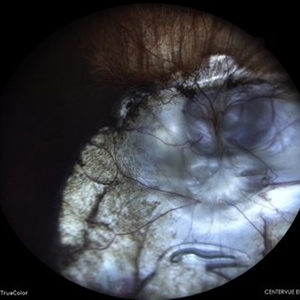
Optic nerve coloboma and rhegmatogenous retinal detachment
Photographer: Píndaro Alonso Cruz-Benitez, APEC, Mexico
Condition/keywords: optic nerve coloboma, Retinal Detachment
-
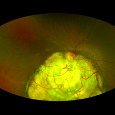
 Double Bubble Coloboma Appearance
Sep 14 2025 by SHADAB HASAN
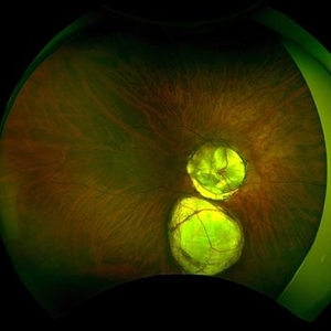
This ultra-widefield fundus photograph shows a large, well-demarcated, whitish excavated involving the inferior fundus with two distinct lobes, giving a “double-bubble” appearance. These features are characteristic of an inferior choroidal coloboma.
Photographer: SHADAB HASAN
Imaging device: OPTOS
Condition/keywords: Coloboma
-
 Posterior Staphyloma + ON-Coloboma
Aug 20 2025 by Gustavo Uriel Fonseca Aguirre
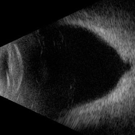
This axial B-scan reveals a highly myopic eye with a posterior staphyloma and an associated optic nerve coloboma. The staphyloma appears as a deep scleral outpouching adjacent to the optic disc, while the coloboma demonstrates a focal posterior excavation with retrobulbar extension.
Photographer: Gustavo U. Fonseca Aguirre, Hospital Conde de Valenciana, Ciudad de México
Condition/keywords: optic nerve coloboma, posterior staphyloma
-
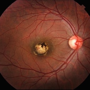
 Presumed Congenital Toxoplasmosis Macular Coloboma
Aug 16 2025 by Vishal Agrawal, MD, FRCS,FACS,FASRS
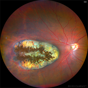
7-year-old boy presented with esotropia in OD with light perception positive. Fundus reveals a large macular coloboma occupying nearly the entire macula. OCT scan shows complete atrophy and disorganization of the overlying RPE and neurosensory retina. A much smaller lesion was observed in OS with BCVA 20/40.
Photographer: Dr Ayushi Gupta
Imaging device: Clarus 700
Condition/keywords: Coloboma, congenital toxoplasmosis
-
 Presumed Congenital Toxoplasmosis
Aug 16 2025 by Vishal Agrawal, MD, FRCS,FACS,FASRS
Fundus picture of 7 a year-old boy with esotropia. OCT showed complete atrophy & disorganization of the overlying RPE and neurosensory retina.
Photographer: Dr Ayushi Gupta
Imaging device: Clarus 700
Condition/keywords: coloboma of macula, toxoplasmosis
-
 Pseudoduplication of the Optic Disc
Jul 9 2025 by Hrishikesh Naik, MS
A peripapillary colobomatous pseudo-duplication of the optic disc as seen in an asymptomatic 23 year old female with myopia referred for routine retinal periphery screening. Rest retinal exam was normal. Duplication of the optic disc can be classified as either true duplication or pseudoduplication, both of which are rare clinical conditions. Pseudodoubling of the optic disc is commonly caused by optic disc or peripapillary colobomas, characterized by a circumscribed, disc-like lesion with radiating vessels but only one normal optic nerve. A few cases have involved pathological myopia, moderate myopia, proliferative diabetic retinopathy and CHARGE syndrome. The lesion is often found inferior to the normal optic disc. The patient was advised regular follow ups.
Photographer: Hrishikesh Naik
Imaging device: Optos Daytona
Condition/keywords: Coloboma, Pseudoduplication of optic disc
-
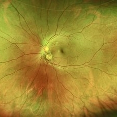
 Coloboma of Optic Disc
Jun 24 2025 by yao zhang
Coloboma of optic disc.
Photographer: Yao Zhang,TongUniversit
Condition/keywords: coloboma of optic disc
-
 Macular Coloboma
Jun 5 2025 by César Adrián Gómez Valdivia, MD
Macular Coloboma found in a 28 year-old male patient, visual acuity was 20/60. Resulting due to fusion failure of the optic fissure, colobomas are commonly found in the infero-nasal quadrant. If the retina is involved, it is reduced to glial tissue with no underlying RPE or choroid. This appears as an area of whitening often with pigment deposition at the junction of the coloboma and normal retina. Findings were bilateral.
Photographer: @eyemissu2
Imaging device: TOPCON TRC-50DX
Condition/keywords: coloboma
-
 Unilateral Coloboma Involving Disc and Macula
Dec 27 2024 by Tejaswita Verma
Fundus image of a 15 years old male presenting with unilaterally diminished vision since childhood in RE with CF3mt vision and inferior iris coloboma and retinochoroidal coloboma with nystagmus and cataract.
Photographer: DR. TEJASWITA VERMA
Imaging device: MIRANTE
Condition/keywords: chorioretinal coloboma, iridofundal coloboma
-
 Coloboma in a Unicameral Eye
Dec 20 2024 by Virginia Gebhart
59 year old female with choroidal coloboma extending into iris. Pt had PC IOL placed in 2016, removed in Aug due to suspected UGH syndrome. Lens haptics were oriented vertically causing haptic to chafe iris superiorly. Most likely etiology was loss of inferior zonules from coloboma. Pt remains aphakic.
Photographer: Virginia Gebhart, Retina Consultants of Carolina
Imaging device: Optos California
Condition/keywords: choroidal coloboma, coloboma
-
 Coloboma
Dec 12 2024 by DANNA ALEXANDRA ALVIRDE AYALA
Ultra-wide field image of a 30-year-old man with coloboma.
Photographer: Danna Alexandra Alvirde-Ayala, Hospital Militar de Especialidades Oftalmológicas, Ciudad de México, México
Imaging device: Optos ultra-widefield
Condition/keywords: coloboma, optic nerve, retina
-
 Optic Disc With Choroidal Coloboma
Nov 9 2024 by LUBNA AHMAD
Optic disc coloboma with isolated fovea sparing choroidal coloboma with stable intercalycial fluid.
Photographer: Shubham
Imaging device: zeiss clarus 500
Condition/keywords: choroidal coloboma
-
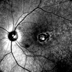 Signet Ring in the Eye
Sep 28 2024 by Tejaswita Verma
Infrared fundus image of the LE of a 32 year-old male showing macular coloboma.
Photographer: DR. TEJASWITA VERMA
Imaging device: MIRANTE
Condition/keywords: macular coloboma
-
 Fundal Coloboma
Sep 25 2024 by DR Rohit Gupta
Fundus photograph of 16year old female patient with a fundal coloboma in left eye
Photographer: Dr Rohit gupta
Imaging device: Samsung S21
Condition/keywords: chorioretinal coloboma, coloboma of macula, coloboma of optic disc, congenital anomaly
-
 OCT in Case of Macular Coloboma (LE)
Sep 18 2024 by Anand Temkar
A 24 year old male came with chief complaint of diminution of vision in both eyes since childhood. Vision in both eyes was 6/24. IOP in RE was 12 and LE was 14 mm of Hg. On fundus examination periphery was within normal limits and central fundus revealed this picture. The serology testing such as serum IgM, IgG for toxoplasma and cytomegalovirus was negative. I have also uploaded LE color photo and BE OCT of this patient.
Photographer: Dr.Anand Temkar- Retina Foundation, Ahmedabad
Imaging device: Mirante
Condition/keywords: Coloboma
-
 OCT in Case of Macular Coloboma (RE)
Sep 18 2024 by Anand Temkar
A 24 year old male came with chief complaint of diminution of vision in both eyes since childhood. Vision in both eyes was 6/24. IOP in RE was 12 and LE was 14 mm of Hg. On fundus examination periphery was within normal limits and central fundus revealed this picture. The serology testing such as serum IgM, IgG for toxoplasma and cytomegalovirus was negative. I have also uploaded LE color photo and BE OCT of this patient.
Photographer: Dr.Anand Temkar- Retina Foundation, Ahmedabad
Imaging device: Mirante
Condition/keywords: coloboma
-
 Macular Coloboma (LE)
Sep 18 2024 by Anand Temkar
A 24 year old male came with chief complaint of diminution of vision in both eyes since childhood. Vision in both eyes was 6/24. IOP in RE was 12 and LE was 14 mm of Hg. On fundus examination periphery was within normal limits and central fundus revealed this picture. The serology testing such as serum IgM, IgG for toxoplasma and cytomegalovirus was negative. I have also uploaded LE color photo and BE OCT of this patient.
Photographer: Dr.Anand Temkar- Retina Foundation, Ahmedabad
Imaging device: Mirante
Condition/keywords: macular coloboma
-
 Macular Coloboma (RE)
Sep 18 2024 by Anand Temkar
A 24 year old male came with chief complaint of diminution of vision in both eyes since childhood. Vision in both eyes was 6/24. IOP in RE was 12 and LE was 14 mm of Hg. On fundus examination periphery was within normal limits and central fundus revealed this picture. The serology testing such as serum IgM, IgG for toxoplasma and cytomegalovirus was negative. I have also uploaded LE color photo and BE OCT of this patient.
Photographer: Dr.Anand Temkar- Retina Foundation, Ahmedabad
Imaging device: Mirante
Condition/keywords: coloboma of macula
-
 Optic Disc Coloboma
Aug 10 2024 by César Adrián Gómez Valdivia, MD
Optic Disc Coloboma found in an 8YO patient. Findings were bilateral. Unlike the morning glory disc, the ODC has no central glial tuft and the disc vasculature is usually normal.
Photographer: @eyemissyou2
Imaging device: Topcon
Condition/keywords: Coloboma, coloboma of optic disc, optic disc
-
 Retinal Colomoba
Jul 21 2024 by César Adrián Gómez Valdivia, MD
Retinal Coloboma found in a female 41 year old patient. Iris, Lens, Ciliary Body, Zonules, Choroid and Retina were involved.
Photographer: Erika Paulina Ornelas Cazares
Imaging device: TOPCON TRC-50DX
Condition/keywords: coloboma
-
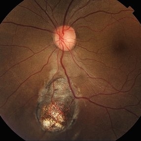
 Macular Coloboma
Jul 17 2024 by Anubhav Chauhan
This is fundus photograph of a 30 year male depicting a Macular coloboma in the right eye. The patient had a sharply defined large, yellowish white, coarsely pigmented, atrophic, round crater like defect at the macula. Spectral domain optical coherence tomography confirmed our diagnosis. The serology testing such as serum IgM, IgG for toxoplasma and cytomegalovirus was negative. His systemic examination was normal.
Photographer: Dr Anubhav Chauhan, Department of Ophthalmology, Shri Lal Bahadur Shastri Government Medical College, Nerchowk, District Mandi, Himachal Pradesh, India
Imaging device: Zeiss
Condition/keywords: macula, rare
-
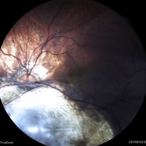 Retinal Detachment With Irido-fundal Coloboma
Feb 7 2024 by Akansha Sharma
Color fundus photograph of a 43 year old male with retinal detachment in a case of iridofundal coloboma.
Photographer: Dr. Akansha Sharma, Bharati Eye Hospital
Condition/keywords: chorioretinal coloboma, RD
-
 Status Post Retinal Detachment Surgery in a Case of Iridofundal Coloboma
Feb 7 2024 by Akansha Sharma
Color fundus photograph of a 43 year old male post retinal detachment surgery in a case of iridofundal coloboma.
Photographer: Dr. Akansha Sharma, Bharati Eye Hospital
Condition/keywords: chorioretinal coloboma, RD
-
 IOL Drop in a Case of Iridofundal Coloboma
Feb 7 2024 by Akansha Sharma
Color fundus photograph of a 43 year old male with IOL drop in a case of iridofundal coloboma.
Photographer: Dr. Akansha Sharma, Bharati Eye Hospital
Condition/keywords: chorioretinal coloboma, IOL drop
 Loading…