Search results (277 results)
-
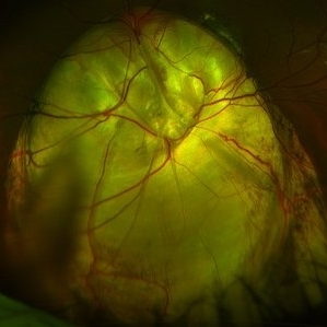
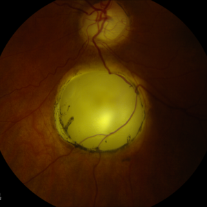 Coloboma
Dec 28 2012 by Carl C. Awh, MD, FASRS
Photographer: Alecia Camp, CRA - Tennessee Retina - Nashville, TN
-
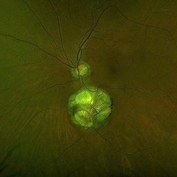
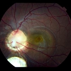
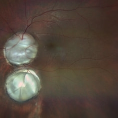
 Optic Disc Coloboma`
Mar 26 2018 by Purva Patwari
16-year-old female patient with vision of 6/60 presented with diminished vison. Other eye was normal.She had a normal birth history and developmental milestone. Look at the optic disc coloboma extending upto the macula. Intercalary membrane looks normal.
Photographer: Dr Purva Patwari, Patwari Retina Center, Ahmedabad, Gujarat , India
Imaging device: ZEISS VISU 500
Condition/keywords: coloboma, coloboma of optic disc, optic disc
-
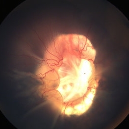 Retinal Detachment Associated with Coloboma
Aug 23 2020 by Noy Ashkenazy, MD, MS
Fundus photograph of a 2-year-old boy with a history of CHARGE syndrome. The image nicely illustrates a retinal detachment associated with a congenital coloboma.
Photographer: Giselle DeOliveira
Imaging device: Retcam III
Condition/keywords: CHARGE syndrome, chronic retinal detachment, coloboma, pediatric retina
-
 Cat Eye Syndrome
Feb 11 2020 by Sophia El Hamichi, MD
A 3-year-old female with cat eye syndrome including iris, chorioretinal and optic nerve colobomas. Note the CNV temporally to the optic nerve coloboma (blue arrows)
Photographer: Giselle De Oliveira, Bascom Palmer Eye Institute, Miami
Imaging device: RetCam
Condition/keywords: cat eye syndrome, chorioretinal coloboma, choroidal neovascularization (CNV), coloboma, coloboma of optic disc, optic nerve coloboma
-
 Macular Coloboma OS
Sep 26 2012 by Jose Dalma-Weiszhausz, MD
25-year-old male with poor vision since birth OU.
Photographer: José Dalma, MD, Dalma & Asoc. Mexico City, Mexico
Condition/keywords: macular coloboma
-
 Presumed Congenital Toxoplasmosis
Aug 16 2025 by Vishal Agrawal, MD, FRCS,FACS,FASRS
Fundus picture of 7 a year-old boy with esotropia. OCT showed complete atrophy & disorganization of the overlying RPE and neurosensory retina.
Photographer: Dr Ayushi Gupta
Imaging device: Clarus 700
Condition/keywords: coloboma of macula, toxoplasmosis
-
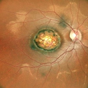
 Chorioretinal Coloboma
Aug 7 2023 by Aditya S Kelkar, MS, FRCS, FASRS,FRCOphth
Fundus photograph of an 68-year-old woman with a chorioretinal coloboma observed.
Photographer: Optom Komal Jangam, National Institute of Ophthalmology, Pune, India.
Imaging device: OPTOS DAYTONA
Condition/keywords: chorioretinal coloboma
-
 Chorioretinal Coloboma with Retinal Detachment
Dec 5 2020 by Niloofar Piri, MD
14-year-old female with 1q21.1 microdeletion syndrome and behavioral, intellectual, and systemic abnormalities, including congenital microcornea, iris coloboma, and chorioretinal and optic nerve coloboma presented with decreased vision. Right eye fundus taken with RetCam shows coloboma with retinal detachment. (Left eye showed white cataract with funnel RD on B-scan).
Photographer: Niloofar Piri MD, Douglas Snyder MD
Condition/keywords: chorioretinal coloboma, optic nerve coloboma
-
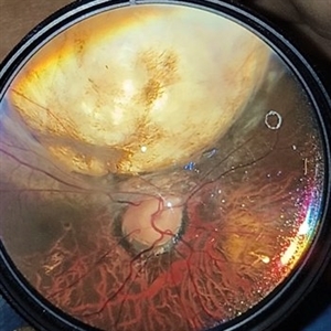
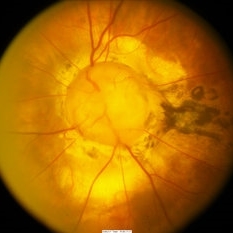
 Coloboma involving the Optic nerve, Retina, and Choroid
Dec 6 2021 by Jesus Lozano, MD
78-year-old woman after prophylactic laser photocoagulation (PLP) for her RE Coloboma involving the optic nerve, retina, and choroid. At 6 month follow up, patient preserved her FC vision as it was before the procedure. Retina attached.
Photographer: Yair Bet Yosef, Hadassah Medical Center. Israel
Imaging device: Optos Silverstone fundus image
Condition/keywords: coloboma, coloboma of choroid, coloboma of macula, coloboma of optic disc, PLP, prophylactic photocoagulation
-
 Coloboma of Optic Disc
Apr 28 2019 by Bastián Schmidt Arias
Fundus photograph of an 63-year-old woman with retinal coloboma.
Photographer: Bastian Schmidt
Condition/keywords: coloboma of optic disc
-
 Colobomatous Optic Disc Maculopathy
Feb 13 2020 by Yoshihiro Yonekawa, MD, FASRS
Beautifully focused fundus photograph of a teenage girl with submacular fluid from a colobomatous optic disc.
Photographer: Netanya Lerner, COA, Wills Eye Hospital/Mid Atlantic Retina
Imaging device: Topcon
Condition/keywords: chorioretinal coloboma, coloboma of optic disc, congenital optic nerve pit, subretinal fluid
-
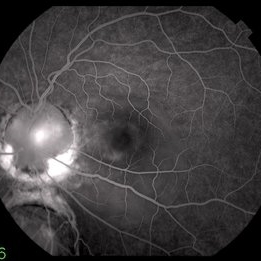 Colobomatous Optic Disc Maculopathy
Feb 13 2020 by Yoshihiro Yonekawa, MD, FASRS
Fluorescein angiography, late frame, of a teenage girl with submacular fluid from a colobomatous optic disc. The camera is focused is on the elevated macula, and the disc is subtly defocused.
Photographer: Netanya Lerner, COA, Wills Eye Hospital/Mid Atlantic Retina
Imaging device: Topcon
Condition/keywords: chorioretinal coloboma, coloboma of optic disc, congenital optic nerve pit, subretinal fluid
-
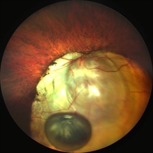 Dislocated Brown Cataract with a Chorioretinal Coloboma
Sep 8 2021 by Ram Sudarshan
A 44 year-old male with dislocated brown cataract along with a chorioretinal coloboma.
Photographer: Dr.Sivadarshan
Condition/keywords: Brown cataract, chorioretinal coloboma, d, dislocated lens
-
 Fundal Coloboma
Mar 6 2023 by Kalyan Singh
34 year old male with fundal Coloboma presented for refractive correction.
Photographer: Kalyan Singh, GSVM medical college, Kanpur
Imaging device: Smartphone (1 plus 10R)
Condition/keywords: coloboma
-
 Fundus Coloboma
Feb 22 2023 by Zach Seim
An ultra-widefield fundus image of a 25 year old male with Fundus Coloboma, as well as Iris Coloboma affecting both eyes. Patient's vision at the time of the image was 20/100-2. Discussed genetic testing as patient reports that he has a child with coloboma and patient agrees. There is a possibility of this finding being syndromic given cornea has small WTW and possibly microphthalmia. The patient has old tractional exudation at edge (abutting fovea). Recommended observation without treatment.
Photographer: Zach Seim
Imaging device: Optos California
Condition/keywords: coloboma, coloboma of optic disc, fundus photograph, Optos, scanning laser ophthalmoscope, ultra-wide field imaging
-
 Inferior Choroidal Coloboma and Tilted Disc
Feb 19 2013 by From the Collections of Thomas M. Aaberg, MD and Thomas M. Aaberg Jr., MD
NLP; Left of stereo pair.
Condition/keywords: coloboma, stereo pair
-
 MACULAR COLOBOMA
Oct 15 2022 by Akansha Sharma
COLOUR FUNDUS PHOTOGRAPH OF A 32 YEAR OLD MALE WITH MACULAR COLOBOMA
Photographer: Dr. Akansha Sharma-Retina Foundation, Ahmedabad
Condition/keywords: coloboma of macula
-
 Macular Coloboma
Jul 17 2024 by Anubhav Chauhan
This is fundus photograph of a 30 year male depicting a Macular coloboma in the right eye. The patient had a sharply defined large, yellowish white, coarsely pigmented, atrophic, round crater like defect at the macula. Spectral domain optical coherence tomography confirmed our diagnosis. The serology testing such as serum IgM, IgG for toxoplasma and cytomegalovirus was negative. His systemic examination was normal.
Photographer: Dr Anubhav Chauhan, Department of Ophthalmology, Shri Lal Bahadur Shastri Government Medical College, Nerchowk, District Mandi, Himachal Pradesh, India
Imaging device: Zeiss
Condition/keywords: macula, rare
-
 Morning Glory Disc Anomaly
Nov 11 2020 by Yoshihiro Yonekawa, MD, FASRS
Color fundus photograph of a young boy with morning glory disc anomaly. Notice the concavity surrounding the enlarged disc, radial vasculature, and nasally dragged macula. MRI was negative for moyamoya disease, a known association.
Photographer: Alicia Thresher, Mid Atlantic Retina
Imaging device: Topcon
Condition/keywords: disc coloboma, Morning Glory Syndrome, pediatric retina
-
 Optic Disc Coloboma
Aug 27 2022 by Aditya S Kelkar, MS, FRCS, FASRS,FRCOphth
Color fundus photograph of a 51-year-old man showing optic disc coloboma of the left eye.
Photographer: Dr. Sukanya Mondal. National Institute of Ophthalmology, Pune, India.
Imaging device: Zeiss Clarus 500
Condition/keywords: coloboma of optic disc, color fundus photograph
-
 Optic Disc Coloboma
Apr 25 2017 by Nimrod Dar
9 year-old patient, noticed a gradual deterioration in her visual acuity at her LE (6/15). On her examination, a double optic disc can be seen. OCT scan revealed an intra retinal fluid and macular schisis.
Photographer: Nimrod Dr, MD
Condition/keywords: coloboma of the optic nerve
-
 Optic Disc Coloboma
Sep 18 2016 by John T. Thompson, MD
Optic disc coloboma
Imaging device: Zeiss FF4
Condition/keywords: coloboma, optic disc
-
 Optic Disc Coloboma
Aug 10 2024 by César Adrián Gómez Valdivia, MD
Optic Disc Coloboma found in an 8YO patient. Findings were bilateral. Unlike the morning glory disc, the ODC has no central glial tuft and the disc vasculature is usually normal.
Photographer: @eyemissyou2
Imaging device: Topcon
Condition/keywords: Coloboma, coloboma of optic disc, optic disc
-
 Optic Disc With Choroidal Coloboma
Nov 9 2024 by LUBNA AHMAD
Optic disc coloboma with isolated fovea sparing choroidal coloboma with stable intercalycial fluid.
Photographer: Shubham
Imaging device: zeiss clarus 500
Condition/keywords: choroidal coloboma
-
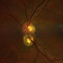
 Pseudo-doubling of optic nerve with Coloboma
Jul 10 2023 by Aditya S Kelkar, MS, FRCS, FASRS,FRCOphth
Right eye fundus photograph of a 41 year old asymptomatic female demonstrating Pseudo-doubling of optic nerve with Coloboma.
Photographer: Miss Komal Jangam,B.Sc Optometry, National Institute of Ophthalmology, Pune, India.
Imaging device: OPTOS DAYTONA
Condition/keywords: coloboma, Pseudoduplication of optic disc
 Loading…