File number: 27949
Comments
-
 Suber S. Huang, MD, MBA, FASRS (April 20 2018)
Focus, exposure, and magnification make this image SUPERB. Great capture by the Patwari team! Please send additional clinical details if possible to improve the quality of the case. Please keep posting and thank you for sharing!
Sign in to comment.
 Initializing download.
-
 By Purva Patwari
Patwari Retina Clinic
Co-author(s): Ruchi Tripathi - Uploaded on Mar 26, 2018.
- Last modified by Caroline Bozell on Jun 12, 2018.
- Image of the week
-
Jun 10, 2018
View all images of the week - Rating
- Appears in
- Patwari Retina Clinic
- Condition/keywords
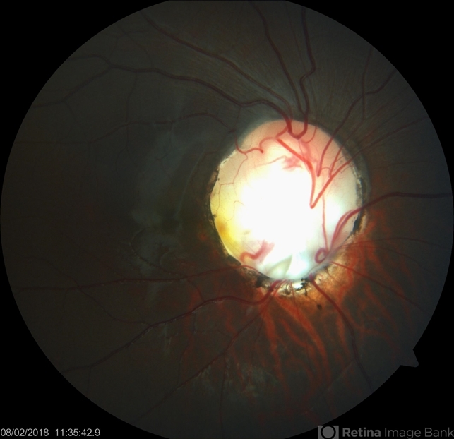
- optic disc, coloboma, coloboma of optic disc
- Photographer
- Dr Purva Patwari, Patwari Retina Center, Ahmedabad, Gujarat , India
- Imaging device
-
Fundus camera
ZEISS VISU 500 - Description
- 16-year-old female patient with vision of 6/60 presented with diminished vison. Other eye was normal.She had a normal birth history and developmental milestone. Look at the optic disc coloboma extending upto the macula. Intercalary membrane looks normal.


---thumb.jpg/image-square;max$79,0.ImageHandler "Coloboma")
---thumb.jpg/image-square;max$79,0.ImageHandler "Melanocytoma")
---thumb.jpg/image-square;max$79,0.ImageHandler "Optic Disc and Retinal Edema")


