Search results (22 results)
-
 CHRPE and Bear Tracks
Jan 7 2025 by Drew Mitchell
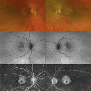
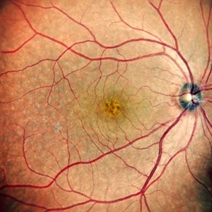
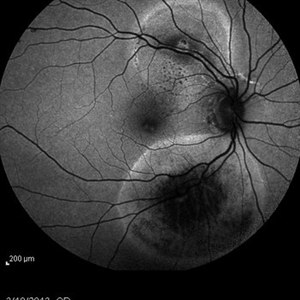
Fundus Autofluorescence of a CHRPE and Bear Tracks.
Photographer: Drew Mitchel, OCT-C
Imaging device: Optos Silverstone
Condition/keywords: bear tracks, CHRPE, congenital hypertrophy of the retinal pigment epithelium (CHRPE)
-
 Acute Syphilitic Posterior Placoid Chorioretinitis
Oct 20 2024 by César Adrián Gómez Valdivia, MD
Fundus autofluorescence image of an acute syphilitic posterior placoid chorioretinitis found in a HIV positive 28 YO male patient with suspected neurosyphilis. A beautiful butterfly autofluorescence pattern can be appreciated.
Photographer: @eyemissu2
Imaging device: California ICG OPTOS
Condition/keywords: acute syphilitic posterior placoid chorioretinitis
-
 Choroidal Metastasis With Orange Pigment in a Patient With Endometrial Carcinoma
Aug 8 2024 by Guilherme Sturzeneker, MD, MSc
Ultra-widefield fundus photograph and autofluorescence of a 62-year-old woman with endometrial cancer, denoting choroidal metastasis with unusual orange pigment. This presentation is a reminder that the development of orange pigment is not pathognomonic for choroidal melanoma, as it may be seen in other lesions such as carcinoma metastasis.
Photographer: Andrea Almeida
Imaging device: Optos Silverstone
Condition/keywords: choroidal metastasis, metastatic cancer, orange pigment
-
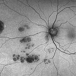
 Pericentral Retinitis Pigmentosa
Sep 6 2024 by Mauricio Bayram-Suverza, MD
A 65-year-old male patient reports experiencing bilateral blind spots that have gradually intensified over time. Genetic testing was unrevealing. The fundus autofluorescence image shows a hypoautofluorescent ring in the posterior pole, especially nasal to the nerve and along arcades.
Photographer: Mauricio Bayram-Suverza, Casey Eye Institute, OHSU.
Imaging device: Optos California
Condition/keywords: fundus autofluorescence (FAF), inherited retinal disease, nyctalopia, retinal dystrophy, retinitis pigmentosa
-
 B-FAF in Stargardt's Disease
Jul 4 2024 by Tejaswita Verma
Blue fundus autofluorescence showing hypoautofluorescence picture of a 28 year old male with 6/60 vision in BE in a case of Stargardt's disease.
Photographer: DR. TEJASWITA VERMA
Imaging device: MIRANTE
Condition/keywords: fundus autofluorescence (FAF), hereditary macular dystrophy, Stargardt disease
-
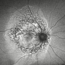
 Rod Cone dystrophy
Nov 29 2022 by Niloofar Piri, MD
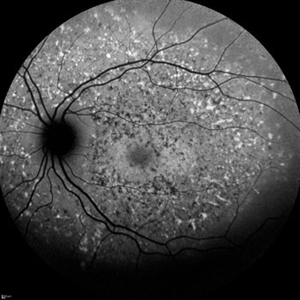
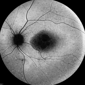
Fundus autofluorescence of the left eye in a 58 yo male with rod cone dystrophy. He presented with night blindness and peripheral vision loss since youth and recent decrease in central vision for the past 10 years. Notice multiple coin shaped hypoautofluorescent pacthes within central 20 degrees which are coalescing centrally. (fundus photo uploaded separately) He has one pathogenic variants of both CEP290 and PRPH2 genes.
Photographer: Sean Kelso, Saint Louis University
Condition/keywords: hereditary retinal degeneration, hereditary retinal dystrophy, rod cone dystrophy
-
 Angioid Streaks
Dec 14 2022 by Pramod Kumar Suman, MBBS, MD
Fundus autofluorescence photograph of a 65-year-old male with numerous narrow, irregular streaks radiating in a circumferential pattern within the posterior pole with macular atrophy.
Photographer: Pramod Kumar Suman, Retina Foundation, Ahmedabad
Imaging device: Mirante
Condition/keywords: angioid streaks
-
 "The Eye of Sauron"
Mar 14 2023 by Anfisa Ayalon, MD
Fundus autofluorescence image of a 38-year-old female with “Bull's eye” pattern maculopathy. There is no history of medication use associated with retinal toxicity. BCVA RE 20/25+2
Photographer: Danielle Ferguson and Alec Bertoni, University of Pittsburgh Medical Center
Condition/keywords: bull's eye maculopathy, retina
-
 Rod cone dystrophy autofluorescence
Sep 19 2022 by Kenneth Fong
34 year old male with colour blindness and loss of visual field
Condition/keywords: retinal dystrophy
-
 CERKL-related Cone Rod Dystrophy
Jun 27 2022 by Hanna Choi
37-year-old female with cone-rod dystrophy. Developed photophobia and progressive blurry vision in the third decade. VA 20/40 OD, 20/30 OS. The patient is compound heterozygous for pathogenic mutations in the CERKL gene (Arg465Trp and Arg283*).
Photographer: Kaitlynn Silva, New England Retina Consultants
Imaging device: Ultrawide-field Optos Fundus Photography, Autofluorescence, Fluorescein Angiography
Condition/keywords: cone dystrophy, inherited retinal disease, maculopathy
-
 Fundus Flavimaculatus
Dec 9 2021 by Filip Kecer
Fundus autofluorescence of a 13-year-old girl with suspected Fundus flavumaculatus.
Photographer: Filip Kecer
Imaging device: Spectralis, Heidelberg Engineering
Condition/keywords: fundus flavimaculatus, Stargardt disease
-
 Stargardt Disease
Dec 9 2021 by Filip Kecer
Fundus autofluorescence of a 14-year-old girl with genetically confirmed Stargardt disease.
Photographer: Filip Kecer
Imaging device: Spectralis, Heidelberg Engineering
Condition/keywords: autofluorescence imaging, Stargardt disease
-
 Puzzle Retinitis
Jan 20 2021 by Jamin S. Brown, MD
Puzzle artifact after imaging on a smaller field of view with blue light autofluorescence.
Photographer: Stefanie Palmer CRA, Retina Vitreous Surgeons of CNY
Condition/keywords: autofluorescence imaging, normal eye
-
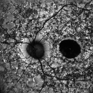
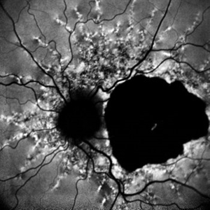
 Optic Disc Drusen and Angioid Streaks
Jun 3 2020 by Mirko Ratkovic, MD
Optic disc drusen and angioid streaks.
Condition/keywords: angioid streaks, fundus autofluorescence (FAF), optic disc drusen
-
 Acute Macular Neuroretinopathy
Dec 11 2019 by Lauren Whaley
34-year-old female patient presented with changes in vision after recent upper respiratory infection. Referring doctor originally thought it was a blood pressure issue. She noticed a "C" shape in her vision. Infrared image was captured showing exactly what patient was describing! Doctor confirmed with this image that it was AMN.
Photographer: Lauren R. Whaley, COA
Imaging device: Heidelberg Spectralis
Condition/keywords: 30 degrees, acute macular neuroretinopathy, Heidelburg Spectralis, left eye, macula, near infrared autofluorescence (NIRAF)
-
 Presumed Tenofovir Induced Toxicity
Nov 7 2019 by Sham Talati, DOMS
46-year-old HIV positive diabetic male with progressive bilateral decrease in vision for last 45 days. Patient had associated liver and kidney disease. Taking Tenofovir for last one year.
Photographer: Sham Talati,Retina Foundation,Ahmedabad
Imaging device: Nidek Mirante
Condition/keywords: autofluorescence imaging, drug toxicity, HIV
-
 Best Disease
Sep 28 2016 by Maciej Czepita
Color fundus image and fundus autofluorescence image of a 41-year-old male patient with Best disease (pseudohypopyon stage).
Photographer: Maciej Czepita, Pomeranian Medical University, Szczecin, Poland
Imaging device: Heidelberg Spectralis HRA+OCT
Condition/keywords: Best disease
-
 Autofluorescence of Retinitis Pigmentosa
Jul 13 2016 by Linda A Cernichiaro- Espinosa, MD
Fundus autofluorescence of an 53-year-old woman with retinitis pigmentosa.
Photographer: Tec Ricardo Montoya, Clínica Oftalmológica Anzures
Condition/keywords: retinitis pigmentosa
-
 RIP 2 FAF
Oct 7 2015 by Roberto Gallego-Pinazo, MD, PhD, DiSSO
Multicolor and autofluorescence sequence of a retinal pigment epithelium tear following intravitreal anti-VEGF injection.
Photographer: Rosa Dolz-Marco, University and Polytechnic Hospital La Fe, Valencia, Spain
Condition/keywords: age-related macular degeneration (AMD), autofluorescence imaging, choroidal neovascularization (CNV), multicolor, retinal pigment epithelium (RPE) tear
-
 Epiretinal Membrane/Macular Pucker With Combined Hamartoma of Retina and RPE
Jul 8 2015 by Emmanuel Chang, MD PhD FACS FASRS
10-year-old with history of progressive severe distortion in the left eye over the past year.
Photographer: Retina and Vitreous of Texas
Imaging device: Heidelberg Autofluorescence
Condition/keywords: combined hamartoma, epiretinal membrane (ERM), retinal pigment epithelium (RPE) hamartoma
-
 Optic Nerve Head Drusen
Feb 12 2015 by Timothy S Fuller, MD
Fundus autofluorescence image of a 34-year-old woman with striking, asymptomatic optic nerve head drusen.
Photographer: Nice Hesse, Texas Retina Associates
Imaging device: Heidelberg Spectralis
Condition/keywords: drusen of optic disc
-
 Retinal Arterial Macroaneurysms
Jan 22 2015 by Darrell E. Baskin, MD
Fundus autofluorescence image of a 60-year-old woman with two retinal arterial macroaneurysms--one recent and one not.
Photographer: Darrell Baskin, Wilford Hall, Lackland Air Force Base, Texas
Imaging device: Heidelberg Spectralis
Condition/keywords: retinal arterial macroaneurysm
 Loading…