Search results (458 results)
-
 Late Stage Stargardt's Disease
Mar 13 2013 by Hamid Ahmadieh, MD
Autofluorescence imaging of the left eye of a 46-year-old man with decreased VA due to advanced Stargardt's disease.
Photographer: Nayereh Hadipoor, Negah Eye Center, Tehran
Imaging device: Heidelberg Spectralis
Condition/keywords: autofluorescence imaging, Stargardt disease
-
 Retinitis Pigmentosa - Fundus Autofluorescence
Sep 20 2014 by Rameez N Hussain, MD
Fundus autofluorescence of retinitis pigmentosa showing hyperautofluorescent rings or foveal hyperautofluorescence.
Photographer: Dr.Rameez N Hussain, MD, Central Imaging Center, Vitreo Retinal Services, Giridhar Eye Institute, Cochin, India
Imaging device: Heidelberg Blue Peak Autofluorescence imaging.
Condition/keywords: bone spicule, cystoid macular edema (CME), fundus autofluorescence (FAF), retinitis pigmentosa
-
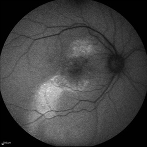
 Serpiginous like Choroiditis
Aug 24 2012 by S. Natarajan, MD, FASRS, FRCS (GLASGOW) , FICO, D.Sc, FELA
Fundus photograph of a 32-year-old male with serpiginous like lesion in the posterior pole followed up with serial autofluorescence imaging.
Photographer: Prof.Dr.S. Natarajan
Imaging device: Zeiss FF 450 plus IR
Condition/keywords: autofluorescence imaging, choroiditis, lesion, serpiginous choroiditis
-
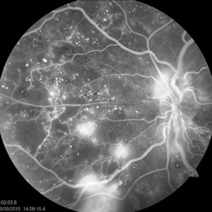
 Retinal Angiomatous Proliferation in Age-Related Macular Degeneration with Subretinal Neovascularization
Sep 24 2012 by James B. Soque, CRA, OCT-C, COA, FOPS
75-year-old white male with classic SRN with RAP. Lesion OD is active, and patient is receiving anti-VEGF treatment. Mid phase FA, 50 Deg, Mag 2x.
Photographer: James Soque, CRA, COA, Island Retina, Shirley, NY, USA
Imaging device: Topcon TRC 50 DX, OIS 5.0 MP Color, FA Camera, OIS Software
Condition/keywords: age-related macular degeneration (AMD), fundus autofluorescence (FAF), leakage, retinal angiomatous proliferation (RAP), subretinal neovascularization (SRNV)
-
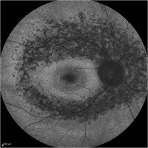
 Stargardts Disease in FAF
Sep 14 2012 by Michael P. Kelly, FOPS
This is a scanning laser ophthalmoscopic FAF image of a patient with Stargardts Disease captured with a Heidelberg Spectralis imaging unit. Note, besides the obvious hyper-autofluorescent areas centrally, the much smaller, and in greater number, pinpoints of hyper-autofluorescence extending from the vascular arcades into the mid-periphery.
Photographer: Michael P. Kelly, FOPS, Director, Duke Eye Center Labs, Duke Universtiy Hospital
Imaging device: Heidelberg Spectralis
Condition/keywords: fundus autofluorescence (FAF), Stargardt disease
-
 Plaquenil Toxicity
Apr 30 2013 by Theodore Leng, MD, MS, FASRS
Fundus autofluorescence from a 44-year-old woman with bilateral plaquenil toxicity. There is an area of hyperautofluorescence that corresponds to areas of outer retinal damage.
Condition/keywords: hydroxychloroquine toxicity, plaquenil toxicity
-
 Vitelliform Macular Dystrophy or Best Disease
Dec 16 2016 by Young Hee Yoon, MD, PhD
Bilateral fundus photographs and autofluorescence images of 15-year-old girl who was diagnosed as vitelliform macular dystrophy or Best disease. Vitelliform macular lesion showed morphologic change during one year.
Photographer: Hyejin Jo, Sunghyun Kim, Heoni Hong, Minjung Chae, Mihwa Shin, Asan medical center, Seoul
Imaging device: Topcon TRC-500X fundus camera, Heidelberg HRA 2 autofluorescence, Heldelberg Spectralis OCT
Condition/keywords: Best disease, pseudohypopyon, scrambled-egg, vitelliform macular dystrophy
-
 Macular Hole, Autofluorescence
Sep 14 2012 by Michael P. Kelly, FOPS
Fundus autofluorescence (FAF) of a macular hole captured using a Heidelberg Spectralis.
Photographer: Michael P. Kelly, FOPS, Director, Duke Eye Cneter Labs, Duke Universty Hospital
Imaging device: Heidelberg Spectralis
Condition/keywords: fundus autofluorescence (FAF), macular hole
-
 Chronic Central Serous Chorioretinopathy re af
Dec 29 2012 by Barbara Parolini, MD
Panoramic autofluorescence fundus photograph of a 56 year old man with chronic central serous chorioretinopathy. BCVA is 20\200.
Photographer: Barbara Parolini, MD
Condition/keywords: bilateral chronic central serous retinopathy
-
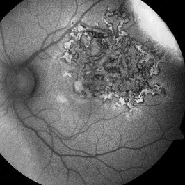
 Epiretinal Membrane/Macular Pucker With Combined Hamartoma of Retina and RPE
Jul 8 2015 by Emmanuel Chang, MD PhD FACS FASRS
10-year-old with history of progressive severe distortion in the left eye over the past year.
Photographer: Retina and Vitreous of Texas
Imaging device: Heidelberg Autofluorescence
Condition/keywords: combined hamartoma, epiretinal membrane (ERM), retinal pigment epithelium (RPE) hamartoma
-
 Plaquenil Toxicity
Apr 30 2013 by Theodore Leng, MD, MS, FASRS
Fundus autofluorescence from a 44-year-old woman with bilateral plaquenil toxicity. There is an area of hyperautofluorescence that corresponds to areas of outer retinal damage.
Condition/keywords: hydroxychloroquine toxicity, plaquenil toxicity
-
Angioid Streaks & CNV (Fig 1)
Aug 25 2012 by Hamid Ahmadieh, MD
Fundus autofluorescence (FAF) of a 53-year-old woman with a juxtafoveal CNV secondary to angioid streaks.
Photographer: Hamid Ahmadieh, Ophthalmic Research Center, Labbafinejad Medical Center
Imaging device: Heidelberg Spectralis
Condition/keywords: angioid streaks, choroidal neovascularization (CNV), fundus autofluorescence (FAF)
-
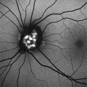
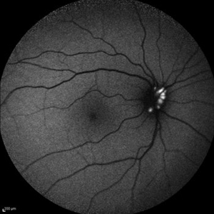 Optic Disc Drusen
Jul 10 2013 by Hamid Ahmadieh, MD
Fundus autofluorescence image of the right eye of a 24-year-old woman with optic disc drusen and VA 20/20.
Photographer: Solmaz Shahmohammadi, Negah Eye Center, Tehran
Imaging device: Heidelberg Spectralis
Condition/keywords: fundus autofluorescence (FAF), optic disc drusen
-
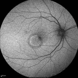 PED due to CSCR 2
Sep 2 2012 by Hamid Ahmadieh, MD
Autofluorescence imaging of a 37-year-old man with a serous PED secondary to CSCR.
Photographer: Hamid Ahmadieh, Ophthalmic Research Center, Labbafinejad Medical Center
Imaging device: Heidelberg Spectralis
Condition/keywords: autofluorescence imaging, central serous chorioretinopathy (CSCR), pigment epithelial detachment (PED)
-
 Multiple Evanescent White Dot Syndrome (MEWDS)
Oct 20 2012 by Hyung-Woo Kwak, MD
Numerous small deep ill-defined, grey-white dot were seen at the posterior pole and mid-periphery. Some lesions showed mild hyperfluorescence in autofluorescence (AF) but were of limited diagnostic value. ICG showed more numerous hypofluorescent spots than are apparent clinically or on AF/FA
Condition/keywords: hypofluorescent spots, multiple evanescent white dot syndrome (MEWDS)
-
 Retinitis Pigmentosa - Autofluorescence OD
Jun 18 2018 by Hosam Attia, MD
Ultra-wide fundus auto-fluorescence photograph of a 38-year-old African, American female with degenerative myopia, unilateral RP variant, depicting extensive mid-peripheral bone spicules hypo-autofluorescence, extending further into the periphery w/ relative sparing of the macula OD VF 30-V showed severe peripheral constriction OD, enlarged BS OS and OCT showed severe ellipsoid zone degeneration with saucerization and cystoid macular degeneration with no obvious late macular leakage on FA (Both, not shown)
Imaging device: Optos California
Condition/keywords: bone spicule, peripheral bone spicules, retinitis pigmentosa
-
 Cystoid Macular Edema (CME)
Sep 11 2012 by Hamid Ahmadieh, MD
Autofluorescence imaging of the left eye of a 17-year-old boy with chronic intermediate uveitis showing CME.
Photographer: Hamid Ahmadieh, MD, Ophthalmic Research Center, Labbafinejad Medical Center, Shahid Beheshti University of Medical Sciences
Imaging device: Heidelberg Spectralis
Condition/keywords: autofluorescence imaging, cystoid macular edema (CME), intermediate uveitis
-
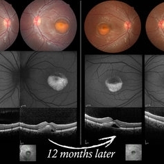
 Hypertensive Choroidopathy - Right Eye
Dec 21 2016 by Maciej Czepita
Fundus photograph and SD-OCT scan as well as fundus autofluorescence image (FAF) of the right eye of a 70-year-old woman with hypertensive choroidopathy. In the fundus image numerous Elschnig's spots are visible. Note the Hollenhorst plaque in the superior temporal artery. In the SD-OCT scan (green line on the fundus image) the RPE layer is uneven. Numerous hypo and hyperautofluorescent patches can be seen in the fundus autofluorescence image.
Photographer: Maciej Czepita, M.D., Ph.D., Pomeranian Medical University, Szczecin, Poland
Imaging device: Heidelberg Spectralis HRA+OCT
Condition/keywords: hypertensive choroidopathy
-
 Multifocal CSCR 1
Sep 2 2012 by Hamid Ahmadieh, MD
Fundus autofluorescence of a 36-year-old man with an active multifocal CSCR.
Photographer: Hamid Ahmadieh, Ophthalmic Research Center, Labbafinejad Medical Center
Imaging device: Heidelberg Spectralis
Condition/keywords: central serous chorioretinopathy (CSCR)
-
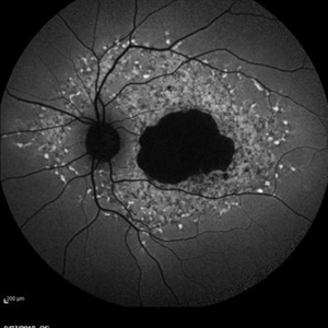
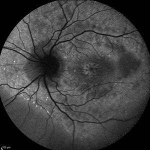
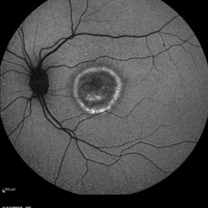
Stargardts Disease in Fundus Autofluorescence
Sep 12 2012 by Michael P. Kelly, FOPS
Fundus autofluorescence of a patient with Stargardts disease. Note the central area of hypo-autofluorescence indicating atrophy surrounded by smaller areas of hyper-autofluorescence. Note also the much smaller, and in greater number, pinpoints of hyper-autofluorescence extending from the vascular arcades into the mid-periphery.
Photographer: Michael P. Kelly, FOPS, Director, Duke Eye Labs, Duke University Hospital, Duke Eye Center
Imaging device: Heidelberg Spectralis
Condition/keywords: fundus autofluorescence (FAF), Stargardt disease
-
 Tamoxifen Retinopathy- FAF
Aug 30 2012 by Young Hee Yoon, MD, PhD
Fundus autofluorescence (FAF) of an 58-year-old woman with a bilateral tamoxifen maculopathy. She had taken tamoxifen for 24 months due to breast cancer. In spite of discontinuation 2 years ago, her macula remained unchanged. Her best-corrected visual acuity was 20/50 in the right and 20/100 in the left.
Photographer: Soo Hyun Cho, Asan Medical Center
Imaging device: Heidelberg HRA II
Condition/keywords: drug toxicity, toxic maculopathy
-
 FFA - PDR
Mar 30 2018 by Lanin Chen
Fundus fluorescein angiography photo of the left eye of a 62-year-old woman with history of Type 2 diabetes mellitus since 20 years showing proliferative diabetic retinopathy.
Photographer: Lanin Chen
Condition/keywords: fundus autofluorescence (FAF), proliferative diabetic retinopathy (PDR)
-
 Optic Disc Drusen Autofluorescence
Apr 2 2016 by David Callanan, MD
30-year-old Caucasian male with visual field defect OD > OS.
Condition/keywords: optic disc drusen
-
 Multiple Evanescent White Dot Syndrome (MEWDS)
Oct 20 2012 by Hyung-Woo Kwak, MD
Numerous small deep ill-defined, grey-white dot were seen at the posterior pole and mid-periphery. Some lesions showed mild hyperfluorescence in autofluorescence (AF) but were of limited diagnostic value. ICG showed more numerous hypofluorescent spots than are apparent clinically or on AF/FA
Condition/keywords: hypofluorescent spots, multiple evanescent white dot syndrome (MEWDS)
-
 Best Disease
Mar 9 2013 by Hamid Ahmadieh, MD
Autofluorescence Imaging of the left eye of a 49-year-old man with decreased VA due to advanced Best disease.
Photographer: Soodabeh Fooladin, Negah Eye Center, Tehran
Imaging device: Heidelberg Spectralis
Condition/keywords: autofluorescence imaging, Best disease
 Loading…