Search results (458 results)
-
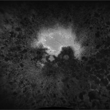
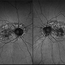
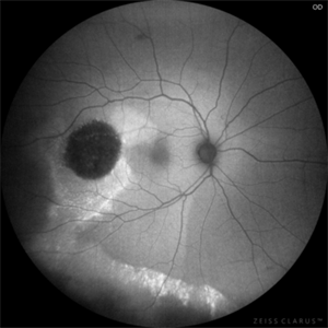
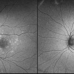
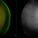 Retinal Macroaneurysm (Left Eye)
Apr 29 2025 by Daniela Bogenschutz
72 year-old female has visual complaints of central vision changes ongoing for 4 days. Patient was acutely symptomatic with an intraretinal hemorrhage due to the retinal macroaneurysm. We had a fun little laugh as this retinal macroaneurysm form a shape of a tick in her left eye. This photo is a side-by-side of the color photos and the autofluorescence done. She is being treated by her general doctor for elevated blood pressure.
Photographer: Daniela Bogenschutz, OSC; Retina Consultants of Carolina, P.A.
Imaging device: Optos
Condition/keywords: retinal macroaneurysm
-
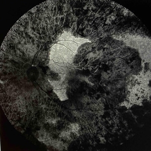 Extensive Chorioretinal Scarring With Partial Macular Sparing
Apr 22 2025 by Maxwell J Wingelaar, MD
Fundus autofluorescence of extensive chorioretinal scarring in the left eye.
Photographer: Killian Roberts
Imaging device: Heidelberg Spectralis AF
Condition/keywords: chorioretinal atrophy, chorioretinal inflammations
-
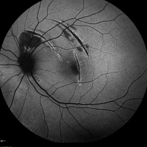
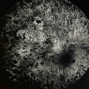 Extensive Chorioretinal Scarring in the Right Eye
Apr 22 2025 by Maxwell J Wingelaar, MD
Fundus autofluorescence of Extensive chorioretinal scarring in the right eye.
Photographer: Killian Roberts
Imaging device: Heidelberg Spectralis AF
Condition/keywords: chorioretinal atrophy, chorioretinal inflammations
-
 Not All Stars Are in the Sky — Some Live in the Eyes of Those Learning to See in New Ways
Apr 21 2025 by rohan jain
Stargardt disease
Photographer: Dr. ROHAN JAIN
Condition/keywords: fleck retinopathy, fundus autofluorescence (FAF), hereditary macular dystrophy
-
 Retinitis Pigmentosa
Apr 17 2025 by Virginia Gebhart
Fundus autofluorescence of 75 year old female with Retinitis Pigmentosa. Pt diagnosed at age 53. Diffuse RPE atrophy with minimal central sparing present in both eyes. Stable and unchanged compared to previous exams. BCVA 20/200 OD, NLP OS
Photographer: Virginia Gebhart, Retina Consultants of Carolina
Imaging device: Optos California
Condition/keywords: autofluorescence imaging, bone spicule, retinitis pigmentosa, RP
-
 Blister Retinal Detachment Superotemporal with a Flap Tear
Apr 10 2025 by Daniela Bogenschutz
Autofluorescence of a 70-year-old male with a superotemporal retinal detachment prior to having an OCT with unusual findings. Patient states symptoms were "starburst" in his vision in the location of the retinal detachment with the retinal tear. Surgery was scheduled immediately to avoid further progression.
Photographer: Daniela Bogenschutz, OSC; Retina Consultants of the Carolinas, PA
Condition/keywords: Retinal Detachment, retinal detachment with single break
-
 LCA Type 2
Apr 10 2025 by Joshua Friedman
LCA Type 2 (RPE65) showing characteristic hypoautofluorescence and retinal thinning. 8F with best corrected visual acuity of 20/400 (OD) and 20/150 (OS). Small white intraretinal spots and RPE mottling are visible on color fundus photography. Blue light autofluorescence reveals near-complete loss of signal, while OCT demonstrates widespread outer retinal thinning.
Photographer: Stephen Tsang, MD, PhD
Condition/keywords: Leber Congenital Amaurosis
-
 LCA type 10
Apr 10 2025 by Joshua Friedman
LCA type 10 due to mutations in CEP290. 36-year-old male with best corrected visual acuity of light perception in both eyes since childhood. On color fundus imaging, there is a mix of polymorphous white flecks and pigmentary changes. On autofluorescence imaging, there is almost complete loss of macular RPE. On OCT, there is complete loss of inner and outer retinal layers, the greatest losses occurring centrally.
Photographer: Stephen Tsang, MD, PhD
Condition/keywords: Leber Congenital Amaurosis
-
 Toxic Maculopathy (Elmiron)
Apr 9 2025 by Virginia Gebhart
79 year old male with toxic maculopathy from long term use of Elmiron (15+ yrs.) On exam there is stippled RPE changes, pigment clumping, and subretinal deposits. BCVA 20/100 | 20/40.
Photographer: Virginia Gebhart, Retina Consultants of Carolina
Imaging device: Optos California
Condition/keywords: autofluorescence imaging, cystoid macular degeneration, Elmiron Toxicity, Toxic Maculopathy
-
 Choroidal Melanoma with Exudative Detachment
Apr 7 2025 by Virginia Gebhart
Autofluorescence image of 36 year old female showing demarcation line of fluid/detachment from new choroidal melanoma. Pt will be scheduled for brachytherapy pending CT scan results.
Photographer: Virginia Gebhart, Retina Consultants of Carolina
Imaging device: Optos California
Condition/keywords: Autoflourescence, autofluorescence imaging, choroidal melanoma, melanoma, retinal detachment
-
 Choroidal Rupture
Apr 7 2025 by Ramses Rosales-Diaz
Autofluorescence image of a 39-year-old female patient who sustained blunt ocular trauma resulting in three choroidal ruptures.
Photographer: Ramses Rosales-Diaz, Asociación Para Evitar la Ceguera en México I.A.P., Mexico City
Imaging device: Heidelberg Spectralis
Condition/keywords: blunt trauma, Choroidal Rupture
-
 Chronic Central Serous Chorioretinopathy (CSCR)
Mar 31 2025 by Niloofar Piri, MD
Fundus Autofluorescence image of the right eye demonstrates classic guttering with hyper autofluorescence consistent with chronic CSCR. Guttering occurs where subretinal fluid migrates inferiorly due to gravity and stressed RPE cells accumulate lipofuscin material from high turnover of photoreceptor outer segments.
Photographer: Stefan Raev, COT; Saint Louis University
Condition/keywords: central serous chorioretinopathy (CSCR), Chronic CSR, Guttering
-
 Elmiron Toxicity
Mar 25 2025 by Toolie Winters
Fundus autofluorescence image of a 69-year-old woman with toxic maculopathy OU due to Elmiron usage. Patient stopped using Elmiron in the late 2010s after having been on it for 17 years. The patient has areas of outer retinal and RPE atrophy temporal to fovea that have expanded compared to photos from two years ago. At the time of this appointment, her VA OD was sc20/40-1+2 PH20/30 and VA OS was scCF @ 1 foot.
Photographer: Toolie Winters
Imaging device: Heidelberg Spectralis
Condition/keywords: Elmiron Toxicity, FAF, fundus autofluorescence (FAF), Heidelburg Spectralis, Pentosan Toxicity, Toxic Maculopathy
-
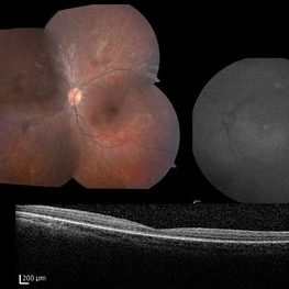
 Choroidal Hemangioma 4 Ways
Mar 13 2025 by Virginia Gebhart
Color fundus, FAF, late FA, late ICG of 64 year old male with choroidal hemangioma. Early hyperfluorescence with late leakage on FA, early hypercyanescence with late washout (25 min) on ICG.
Photographer: Virginia Gebhart, Retina Consultants of Carolina
Imaging device: Optos California
Condition/keywords: autofluorescence imaging, choroidal hemangioma, FA late phase, Fluorescein angiography, hemangioma, indocyanine green (ICG) angiography
-
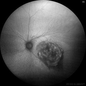 Choroidal Hemangioma, Autofluorescence
Mar 11 2025 by Gustavo Uriel Fonseca Aguirre
45-year-old female with choroidal hemangioma in the macular area. An autofluorescence image of the lesion is presented, with heterogeneous hypoautofluorescent characteristics.
Photographer: Gustavo U. Fonseca Aguirre, Hospital Conde de Valenciana, Ciudad de México
Condition/keywords: Choroidal Hemangioma
-
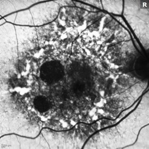
 Posterior Placoid Chorioretinitis
Mar 9 2025 by Oscar Francisco Miranda, MD
A 36-year-old male with bilateral visual loss of 3 months' duration, with no relevant medical history on inquiry. A round-shaped lesion with well-defined borders and a yellowish-white color is observed in the macula of both eyes, accompanied by vitreous cellularity. The macular OCT shows a dentate RPE. The VDRL, FTA-ABS, and HIV tests were positive.
Photographer: Oscar Francisco Miranda-Gómez
Imaging device: Autofluorescence Zeiss Clarus 700
Condition/keywords: acute posterior placoid chorioretinitis, Autofluorescence, ocular syphilis
-
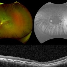
 Multimodal Imaging in CHRPE
Mar 6 2025 by Gerardo - Montante Montelongo, MD
Fundus photograph of an 83-year-old male with a history of Diabetes, smoking, cataract surgery on the right eye in 2022, and open-angle glaucoma. Asymptomatic. Indirect ophthalmoscopy revealed 80% excavation, peripapillary atrophy, and a hyperpigmented perifoveal lesion with 35% atrophy, 10% drusen, and 5.1 mm diameter, corresponding to a CHRPE. At multimodal imaging, FFA shows hypoautofluorescence of the lesion, OCT shows preservation of internal retinal layers, atrophy of external retinal layer, with an RPE disruption, and posterior shadowing. USG shows a flat hyperechoic lesion 5.1 mm in diameter and 1.32 mm in thickness, solid and with high internal reflectance.
Photographer: Gerardo Montante-Montelongo, MD, Mexican Institute of Ophthalmology
Imaging device: Clarus 700
Condition/keywords: congenital hypertrophy of the retinal pigment epithelium (CHRPE), multimodal imaging
-
 Hemangioma of Retina (FAF)
Mar 5 2025 by Virginia Gebhart
Fundus autofluorescence of 64 year old male with choroidal hemangioma in the macula and STA. Persistent IRF and new cuff of SRF compared to previous photos. BCVA CF@face. Pt has had PDT in the past with no significant improvement. Will observe closely
Photographer: Virginia Gebhart, Retina Consultants of Carolina
Imaging device: Optos California
Condition/keywords: autofluorescence imaging, hemangioma, inferior subretinal fluid
-
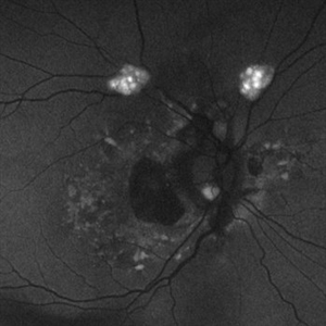
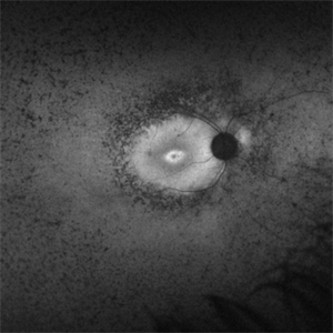
 FAF-G Circumscribed Choroidal Hemangioma
Mar 1 2025 by Vishal Agrawal, MD, FRCS,FACS,FASRS
A 37-year-old male presented with decreased vision in the right eye. This is the fundus autofluorescence (FAF-G) of the right eye showing hypo auto fluorescent lesion with surrounding hyper auto fluorescence extending inferiorly corresponding to the fluid tract.
Photographer: Dr Ayushi Gupta
Imaging device: Clarus 700
Condition/keywords: Circumscribed Choroidal Hemangioma, fundus autofluorescence (FAF)
-
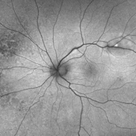
 Rod Cone Dystrophy
Mar 1 2025 by Aditya S Kelkar, MS, FRCS, FASRS,FRCOphth
Fundus Autofluorescence photograph of a 72-year-old woman with a rod cone dystrophy.
Photographer: Optom Rutuja Shelke
Imaging device: OPTOS DAYTONA
Condition/keywords: dystrophy
-
 Astrocytic Hamartoma
Feb 27 2025 by Daniel Davis, OCT-C
Fundus autofluorescence photo of 55-year-old female with astrocytic hamartoma in association with tuberous sclerosis. No treatment options available, benign. Other findings include; Posterior Vitreous Detachment, Vitreous Hemorrhage, Hereditary Retinal Dystrophy, Vitreous Opacities, Hypertensive Retinopathy.
Photographer: Daniel Davis, OCT-C
Imaging device: Optos California
Condition/keywords: astrocytic hamartoma, fundus autofluorescence (FAF)
-
 Resolved Multiple Evanescent White Dot Syndrome
Feb 18 2025 by Jordyn Beckman
Autofluorescence fundus photographs of Resolved Multiple Evanescent White Dot Syndrome in a 28 year old female with previous hyperfluorescent punctate spots throughout the posterior pole.
Photographer: Jordyn Beckman, Retina Consultants of Carolina, P.A.
Imaging device: California Optos
Condition/keywords: autofluorescence imaging, grey-white lesions, multiple evanescent white dot syndrome (MEWDS), scattered punctate
-
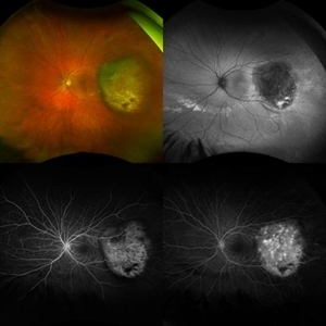 Melanoma Multimodal Evaluation
Feb 10 2025 by Gustavo M. Hüning, MD, MBA, FASRS
UWF multimodal imaging of an 37-year-old woman with a choroidal melanoma. The mosaic shows a colored retinography; a FAF with regions of previous serous detachments; an early stage of angiography and a later time.
Photographer: Gustavo M. Hüning, HÜNING Clínica do Olhar, Santa Maria - Brazil
Imaging device: Optos California
Condition/keywords: Autofluorescence, Choroidal, Fluorescein angiography, melanoma, multimodal imaging, ultra-wide field imaging
-
 Mac-on Retinal Detachment (Barely!)
Feb 6 2025 by Virginia Gebhart
FAF of 46 year old male with a mac-on retinal detachment from 1:00 to 6:00 with a single break at 3:00. Pt scheduled for emergent PPV/Laser/GFE
Photographer: Virginia Gebhart, Retina Consultants of Carolina
Imaging device: Optos California
Condition/keywords: autofluorescence imaging, retinal detachment
-
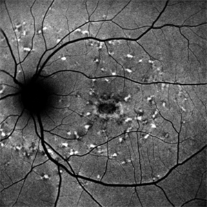
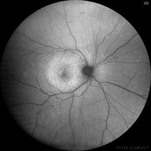
 Retinitis Pigmentosa Bullseye Appearing Autofluorescence
Feb 4 2025 by Isaac Agranoff
Fundus Autofluorescence of a 14-year-old boy with suspected RP. ERG performed afterwards was almost flat. VA measured at 20/30 but with extensive constriction of confrontational visual fields. Currently awaiting genetic testing.
Photographer: Isaac Agranoff
Imaging device: Optos California
Condition/keywords: fundus autofluorescence (FAF), retinitis pigmentosa, RP
 Loading…