Search results (125 results)
-
 Bilateral Papilledema
Jul 14 2013 by Jason S. Calhoun
Patient that had loss of vision and intra-cranial pressure shows papilledema in both eyes.
Photographer: Jason S. Calhoun, Department of Ophthalmology, Mayo Clinic Jacksonville, Florida
Imaging device: TOPCON TRC 50-EX
Condition/keywords: papilledema
-
---thumb.JPG/image-square;max$300,300.ImageHandler) Bilateral Papilledema
Bilateral Papilledema
Jul 14 2013 by Jason S. Calhoun
Patient that had loss of vision and intra-cranial pressure shows papilledema in both eyes.
Photographer: Jason S. Calhoun, Department of Ophthalmology, Mayo Clinic Jacksonville, Florida
Condition/keywords: papilledema
-
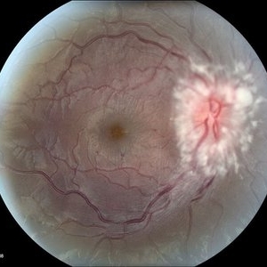
 Biopsy Proven Giant Cell Arteritis
Oct 15 2018 by Darin R. Goldman, MD
83-year-old male with biopsy-proven giant cell arteritis OU and old BRVO OS.
Photographer: Crystal Esparza, BS, COA, Retina Group of Florida
Imaging device: Topcon TRC 50DX
Condition/keywords: branch retinal vein occlusion (BRVO), giant cell arteritis, optic disc edema, papilledema
-
 Chronic Papilledema
Feb 20 2013 by From the Collections of Thomas M. Aaberg, MD and Thomas M. Aaberg Jr., MD
Secondary to cystic _______ of the optic nerve secondary to arachnoiditis.
Condition/keywords: papilledema
-
 Hypertensive Retinopathy
Aug 24 2012 by Geoffrey G. Emerson, MD, PhD, FASRS
A 35-year-old man has headaches and decreased vision. The right eye measures 20/25 and the left eye measures 3/200. The blood pressure measures 180/110. This fluorescein angiogram shows dilated capillaries and capillary dropout in the central macula of the left eye.
Photographer: Geoffrey Emerson, MD, PhD, Retina Center, Minneapolis
Condition/keywords: hypertensive retinopathy, papilledema, retinal ischemia
-
 Hypertensive Retinopathy
Aug 24 2012 by Geoffrey G. Emerson, MD, PhD, FASRS
A 35-year-old man has headaches and decreased vision. The right eye measures 20/25 and the left eye measures 3/200. The blood pressure measures 180/110.This fluorescein angiogram shows leakage of dye from the optic disc (papilledema), ischemia, and dilated capillaries around the foveal avascular zone
Photographer: Geoffrey Emerson, MD, PhD, Retina Center, Minneapolis
Condition/keywords: hypertensive retinopathy, ischemia, papilledema
-
 Hypertensive Retinopathy
Aug 24 2012 by Geoffrey G. Emerson, MD, PhD, FASRS
A 35-year-old man has headaches and decreased vision. The right eye measures 20/25 and the left eye measures 3/200. The blood pressure measures 180/110.
Photographer: Geoffrey Emerson, MD, PhD, Retina Center, Minneapolis
Condition/keywords: hypertensive retinopathy, papilledema, serous retinal detachment
-
 Hypertensive retinopathy
Aug 24 2012 by Geoffrey G. Emerson, MD, PhD, FASRS
A 35-year-old man has headaches and decreased vision. The right eye measures 20/25 and the left eye measures 3/200. The blood pressure measures 180/110.
Photographer: Geoffrey Emerson, MD, PhD, Retina Center, Minneapolis
Condition/keywords: cotton wool spots, hypertensive retinopathy, papilledema
-
 Idiopathic Papillophlebitis- Fluorescein Angiography
Apr 5 2017 by Linda A Cernichiaro- Espinosa, MD
18-year-old otherwise healthy female with sudden visual loss on the left eye.
Photographer: Linda A Cernichiaro MD
Imaging device: Optos Daytona
Condition/keywords: cystoid macular edema (CME), papilledema, venous tortuosity
-
---thumb.jpg/image-square;max$300,300.ImageHandler) Lyme Disease
Lyme Disease
May 20 2013 by Howard Schatz, MD
36-year-old white female, III Lyme disease (papilledema), right eye: 20/20; left eye: 20/16.
Condition/keywords: Lyme disease, papilledema
-
 Mild Patton's Lines in IIH - Initial Photo
Jan 16 2019 by John S. King, MD
18-year-old African American female with increased BMI with a history of headaches, nausea, transient diplopia and vision loss that she notices when getting up from her bed (and goes away after standing upright) for the last two weeks. Went to PCP and was treated for the flu, and after no improvement and visual symptoms known, was sent to ED. MRI did not show any masses and showed empty sella turcia. Vision 20/30 OD and 20/20 OS; no RAPD; IOP 15OU; no anterior segment or vitreous inflammation; discs are elevated with obscuration of the disc margins and some of the smaller vessels; there are no SVPs; there are mild Patton's lines temporally (see Initial Photos). The optic disc cube shows 360 degrees of RNFL thickening (see OCT). Was referred to near-ophthalmologist, Dr. Doyle. She obtained additional work-up, and LP opening pressure was high, and MRV showed bilateral transverse sinus stenosis. Patient showed steady improvement with medical therapy, that included weight loss and oral diamox. On her last visit with Dr. Doyle, vision has remained stable at 20/20-20/25 without an enlarged blindspot; there are SVPs and optic disc edema has resolved (see Post Treatment Photos); she is currently on 1000 mg of diamox and has lost 15 pounds, and no stinting procedure needed.
Photographer: Gretchen Harper
Imaging device: Topcon 50
Condition/keywords: idiopathic intracranial hypertension, optic disc edema, papilledema, Patton's Lines
-
 Mild Patton's Lines in IIH - Initial Photos
Jan 16 2019 by John S. King, MD
18-year-old African American female with increased BMI with a history of headaches, nausea, transient diplopia and vision loss that she notices when getting up from her bed (and goes away after standing upright) for the last two weeks. Went to PCP and was treated for the flu, and after no improvement and visual symptoms known, was sent to ED. MRI did not show any masses and showed empty sella turcia. Vision 20/30 OD and 20/20 OS; no RAPD; IOP 15OU; no anterior segment or vitreous inflammation; discs are elevated with obscuration of the disc margins and some of the smaller vessels; there are no SVPs; there are mild Patton's lines temporally (see Initial Photos). The optic disc cube shows 360 degrees of RNFL thickening (see OCT). Was referred to near-ophthalmologist, Dr. Doyle. She obtained additional work-up, and LP opening pressure was high, and MRV showed bilateral transverse sinus stenosis. Patient showed steady improvement with medical therapy, that included weight loss and oral diamox. On her last visit with Dr. Doyle, vision has remained stable at 20/20-20/25 without an enlarged blindspot; there are SVPs and optic disc edema has resolved (see Post Treatment Photos); she is currently on 1000 mg of diamox and has lost 15 pounds, and no stinting procedure needed.
Photographer: Gretchen Harper
Imaging device: Topcon 50
Condition/keywords: idiopathic intracranial hypertension, optic disc edema, papilledema, Patton's Lines
-
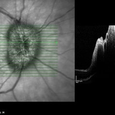
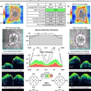 OCT in Patient With IIH Showing Thickened RNFL
Jan 16 2019 by John S. King, MD
18-year-old African American female with increased BMI with a history of headaches, nausea, transient diplopia and vision loss that she notices when getting up from her bed (and goes away after standing upright) for the last two weeks. Went to PCP and was treated for the flu, and after no improvement and visual symptoms known, was sent to ED. MRI did not show any masses and showed empty sella turcia. Vision 20/30 OD and 20/20 OS; no RAPD; IOP 15OU; no anterior segment or vitreous inflammation; discs are elevated with obscuration of the disc margins and some of the smaller vessels; there are no SVPs; there are mild Patton's lines temporally (see Initial Photos). The optic disc cube shows 360 degrees of RNFL thickening (see OCT). Was referred to near-ophthalmologist, Dr. Doyle. She obtained additional work-up, and LP opening pressure was high, and MRV showed bilateral transverse sinus stenosis. Patient showed steady improvement with medical therapy, that included weight loss and oral diamox. On her last visit with Dr. Doyle, vision has remained stable at 20/20-20/25 without an enlarged blindspot; there are SVPs and optic disc edema has resolved (see Post Treatment Photos); she is currently on 1000 mg of diamox and has lost 15 pounds, and no stinting procedure needed.
Imaging device: Cirrus
Condition/keywords: benign idiopatic intracranial hypertension, optic disc edema, papilledema
-
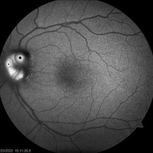
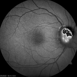 ONH-drusen-OD
Mar 24 2022 by Elite Bor-Shavit, MD
Fundus autofluorescence of a 41-years old patient with combined true papilledema and optic nerve head drusen, treated with Diamox and monitored.
Condition/keywords: optic disc drusen, papilledema
-
 ONH-drusen-OS
Mar 24 2022 by Elite Bor-Shavit, MD
Fundus autofluorescence of a 41-years old patient with combined true papilledema and optic nerve head drusen, treated with Diamox and monitored.
Condition/keywords: optic disc drusen, papilledema
-
---thumb.jpg/image-square;max$300,300.ImageHandler) Optic Disc and Retinal Edema
Optic Disc and Retinal Edema
Feb 13 2013 by From the Collections of Thomas M. Aaberg, MD and Thomas M. Aaberg Jr., MD
Intra-retinal hemorrhage papilledema.
Condition/keywords: intraretinal hemorrhage, optic disc, papilledema, retinal edema
-
 Papiledema
Dec 19 2019 by Zura Glonti
OS. Fundus photograph of 25-years-old man with papilledema, caused by meningioma.
Photographer: Zurab Glonti
Condition/keywords: meningioma, papilledema
-
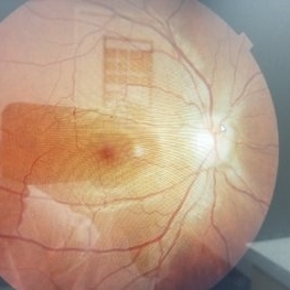 Papilledema
Jan 22 2021 by AGNES KIM
Fundus photograph of 22-year-old woman with papilledema in right eye only. OS ruled out.
Condition/keywords: papilledema
-
 Papilledema
Apr 23 2021 by Amin Masjedi
Fundus photograph of a 32-year-old female with IIH.
Photographer: Amin Masjedi,MD
Condition/keywords: papilledema
-
 Papilledema
Dec 19 2019 by Zura Glonti
OD. Fundus photograph of 25-year-old man with papilledema, caused by meningioma.
Photographer: Zurab Glonti
Condition/keywords: meningioma, papilledema
-
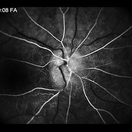
 Papilledema
Feb 20 2013 by From the Collections of Thomas M. Aaberg, MD and Thomas M. Aaberg Jr., MD
Papilledema Fluorescein angiogram
Condition/keywords: papilledema
-
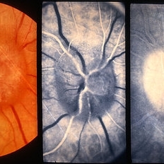
 Papilledema
Feb 20 2013 by From the Collections of Thomas M. Aaberg, MD and Thomas M. Aaberg Jr., MD
Optic nerve head papilledema Fundus photograph
Condition/keywords: optic nerve head, papilledema
-
---thumb.jpg/image-square;max$300,300.ImageHandler) Papilledema
Papilledema
Feb 20 2013 by From the Collections of Thomas M. Aaberg, MD and Thomas M. Aaberg Jr., MD
papilledema
Condition/keywords: papilledema
-
 Papilledema
Nov 21 2016 by JEFFERSON R SOUSA, Tecg.º (Biomedical Systems Technology)
Patient female, 51-year-old, white. attended the clinic with the complaint of low vision, flash, right in the eye and headaches. In the examination of the fundus of the eye were observed important changes such as the Papilledema unilateral. Just confirmation more detailed examination angiographic.
Photographer: JEFFERSON R SOUSA - Study Center and Ophthalmological Research Dr. Andre M V Gomes, Institute Dr. Suel Abujamra São Paulo-Brazil
Imaging device: Topcon TRC-50 Ex - Angulation of field photo of 35 Degrees. Digital system OphthaVision
Condition/keywords: papilledema
-
 Papilledema
Sep 8 2012 by Hamid Ahmadieh, MD
OCT of the optic nerve head of the right eye of a 55-year-old woman with a malignant intracranial tumor.
Photographer: Hamid Ahmadieh, MD, Ophthalmic Research Center, Labbafinejad Medical Center, Shahid Beheshti University of Medical Sciences
Imaging device: Heidelberg Spectralis
Condition/keywords: malignant intracranial tumor, optical coherence tomography (OCT), papilledema
 Loading…